The SPECC1L Antibody (CAB15798) is a high-quality antibody developed for reliable detection and analysis of target proteins. This antibody, produced in rabbits, exhibits high reactivity with human samples and has been validated for use in various applications including Western blot analysis. By targeting the SPECC1L protein, this antibody enables precise detection and analysis in a wide range of cell types, making it an essential component for research in fields such as cellular biology and developmental genetics.
This antibody is validated for use in WB, IHC-P, ELISA applications and has demonstrated reactivity against Human samples.
Product Name:
SPECC1L Antibody
SKU:
CAB15798
Size:
20μL, 100μL
Reactivity:
Human
Conjugate:
Unconjugated
Immunogen:
Recombinant protein (or fragment).This information is considered to be commercially sensitive.
Recommended starting concentration is 1 μg/mL. Please optimize the concentration based on your specific assay requirements.
Synonyms:
TBHS, CYTSA, GBBB2, TBHS1, OBLFC1, SPECC1L
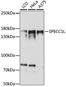
Positive Sample:
LO2, HeLa, A375
Cellular Localization:
Cell Junction, Cytoplasm, Cytoskeleton, Gap Junction, Spindle.
Calculated MW:
125kDa
Observed MW:
168kDa
This gene encodes a coiled-coil domain containing protein. The encoded protein may play a critical role in actin-cytoskeletal reorganization during facial morphogenesis. Mutations in this gene are a cause of oblique facial clefting-1. Alternatively spliced transcript variants encoding multiple isoforms have been observed for this gene. A read-through transcript composed of SPECC1L (sperm antigen with calponin homology and coiled-coil domains 1-like) and the downstream ADORA2A (adenosine A2a receptor) gene sequence has been identified, but it is thought to be non-coding.
Purification Method
Affinity purification
Gene ID
23384
RRID
AB_2763219
Buffer Information
Store at -20℃. Avoid freeze / thaw cycles. Buffer: PBS with 0.01% thimerosal,50% glycerol,pH7.3.
Western blot analysis of various lysates using SPECC1L Rabbit pAb (CAB15798) at 1:1000 dilution. Secondary antibody: HRP-conjugated Goat anti-Rabbit IgG (H+L) (CABS014) at 1:10000 dilution. Lysates/proteins: 25μg per lane. Blocking buffer: 3% nonfat dry milk in TBST. Detection: ECL Basic Kit (AbGn00020). Exposure time: 15s.
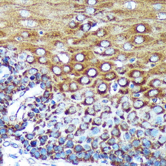
Immunohistochemistry analysis of paraffin-embedded Human esophageal using SPECC1L Rabbit pAb (CAB15798) at dilution of 1:100 (40x lens). Microwave antigen retrieval performed with 0.01M PBS Buffer (pH 7.2) prior to IHC staining.