The SPHK1 Antibody (CAB0139) is a high-quality antibody developed for reliable detection and analysis of target proteins. This antibody, generated in rabbits, is highly specific for human samples and has been validated for use in Western blot applications. By binding to the SPHK1 protein, this antibody enables the detection and analysis of SPHK1 in various cell types, making it an essential tool for studies in immunology, cancer research, and other fields where sphingolipid metabolism is involved.
This antibody is validated for use in WB, ELISA applications and has demonstrated reactivity against Human, Mouse samples.
Product Name:
SPHK1 Antibody
SKU:
CAB0139
Size:
20μL, 100μL
Reactivity:
Human, Mouse
Conjugate:
Unconjugated
Immunogen:
Recombinant protein (or fragment).This information is considered to be commercially sensitive.
Recommended starting concentration is 1 μg/mL. Please optimize the concentration based on your specific assay requirements.
Synonyms:
SPHK, SPHK1
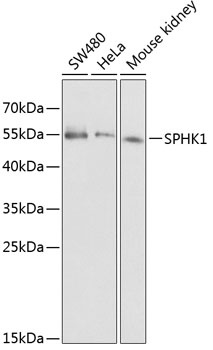
Positive Sample:
SW480, HeLa, Mouse kidney
Cellular Localization:
Cell Membrane, Cytoplasm, Nucleus.
Calculated MW:
43kDa
Observed MW:
55kDa
The protein encoded by this gene catalyzes the phosphorylation of sphingosine to form sphingosine-1-phosphate (S1P), a lipid mediator with both intra- and extracellular functions. Intracellularly, S1P regulates proliferation and survival, and extracellularly, it is a ligand for cell surface G protein-coupled receptors. This protein, and its product S1P, play a key role in TNF-alpha signaling and the NF-kappa-B activation pathway important in inflammatory, antiapoptotic, and immune processes. Phosphorylation of this protein alters its catalytic activity and promotes its translocation to the plasma membrane. Alternative splicing results in multiple transcript variants encoding different isoforms.
Purification Method
Affinity purification
Gene ID
8877
RRID
AB_2756973
Buffer Information
Store at -20℃. Avoid freeze / thaw cycles. Buffer: PBS containing 50% glycerol, preserved with proclin300 or sodium azide, pH 7.3.
Western blot analysis of various lysates using SPHK1 Rabbit pAb (CAB0139) at 1:1000 dilution. Secondary antibody: HRP-conjugated Goat anti-Rabbit IgG (H+L) (CABS014) at 1:10000 dilution. Lysates/proteins: 25μg per lane. Blocking buffer: 3% nonfat dry milk in TBST.