The SQS/FDFT1 Antibody (CAB6229) is a high-quality antibody developed for reliable detection and analysis of target proteins. This antibody, raised in rabbits, is highly specific for human squalene synthase and has been validated for use in Western blot applications.Squalene synthase plays a crucial role in the cholesterol biosynthetic pathway, making it a promising target for therapies aimed at treating dyslipidemia and other related disorders. By detecting and analyzing squalene synthase expression in various cell types, this antibody can provide insights into the regulation of cholesterol synthesis and potential drug targets for cardiovascular diseases and metabolic disorders.
This antibody is validated for use in WB, IHC-P, IF/ICC, ELISA applications and has demonstrated reactivity against Human, Mouse, Rat samples.
Product Name:
SQS/FDFT1 Antibody
SKU:
CAB6229
Size:
20μL, 100μL
Reactivity:
Human, Mouse, Rat
Conjugate:
Unconjugated
Immunogen:
Recombinant protein (or fragment).This information is considered to be commercially sensitive.
This gene encodes a membrane-associated enzyme located at a branch point in the mevalonate pathway. The encoded protein is the first specific enzyme in cholesterol biosynthesis, catalyzing the dimerization of two molecules of farnesyl diphosphate in a two-step reaction to form squalene.
Purification Method
Affinity purification
Gene ID
2222
RRID
AB_2766838
Buffer Information
Store at -20℃. Avoid freeze / thaw cycles. Buffer: PBS containing 50% glycerol, preserved with proclin300 or sodium azide, pH 7.3.
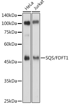
Western blot analysis of various lysates using SQS/FDFT1 Rabbit pAb (CAB6229) at 1:500 dilution. Secondary antibody: HRP-conjugated Goat anti-Rabbit IgG (H+L) (CABS014) at 1:10000 dilution. Lysates/proteins: 25μg per lane. Blocking buffer: 3% nonfat dry milk in TBST. Detection: ECL Enhanced Kit (AbGn00021). Exposure time: 1s.
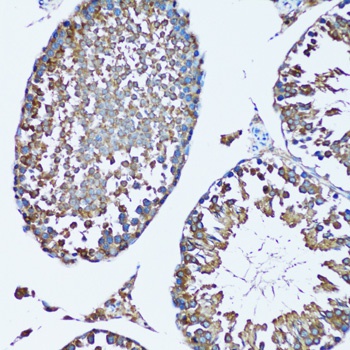
Immunohistochemistry analysis of paraffin-embedded Mouse testis using SQS/FDFT1 Rabbit pAb (CAB6229) at dilution of 1:100 (20x lens). Microwave antigen retrieval performed with 0.01M PBS Buffer (pH 7.2) prior to IHC staining.
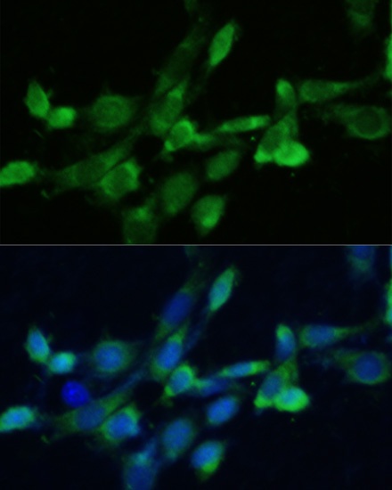
Immunofluorescence analysis of NIH-3T3 cells using SQS/FDFT1 Rabbit pAb (CAB6229) at dilution of 1:100. Secondary antibody: Cy3-conjugated Goat anti-Rabbit IgG (H+L) (CABS007) at 1:500 dilution. Blue: DAPI for nuclear staining.