The SRF Antibody (CAB7731) is a high-quality antibody developed for reliable detection and analysis of target proteins. This antibody, generated in rabbits, exhibits high specificity for human samples and has been validated for use in Western blot applications. By targeting the SRF protein, this antibody allows for accurate detection and analysis in a variety of cell types, making it an essential component for studies in cell biology, developmental biology, and cancer research.SRF is a key player in various cellular processes, including muscle development, neuronal growth, and wound healing.
This antibody is validated for use in WB, IHC-P, ELISA applications and has demonstrated reactivity against Human, Mouse, Rat samples.
Product Name:
SRF Antibody
SKU:
CAB7731
Size:
20μL, 100μL
Reactivity:
Human, Mouse, Rat
Conjugate:
Unconjugated
Immunogen:
Synthetic peptide. This information is considered to be commercially sensitive.
Recommended starting concentration is 1 μg/mL. Please optimize the concentration based on your specific assay requirements.
Synonyms:
MCM1, SRF
Positive Sample:
A-431, NIH/3T3, C6
Cellular Localization:
Nucleus.
Calculated MW:
52kDa
Observed MW:
67kDa
This gene encodes a ubiquitous nuclear protein that stimulates both cell proliferation and differentiation. It is a member of the MADS (MCM1, Agamous, Deficiens, and SRF) box superfamily of transcription factors. This protein binds to the serum response element (SRE) in the promoter region of target genes. This protein regulates the activity of many immediate-early genes, for example c-fos, and thereby participates in cell cycle regulation, apoptosis, cell growth, and cell differentiation. This gene is the downstream target of many pathways; for example, the mitogen-activated protein kinase pathway (MAPK) that acts through the ternary complex factors (TCFs). Two transcript variants encoding different isoforms have been found for this gene.
Purification Method
Affinity purification
Gene ID
6722
RRID
AB_2772384
Buffer Information
Store at -20℃. Avoid freeze / thaw cycles. Buffer: PBS containing 50% glycerol, preserved with proclin300 or sodium azide, pH 7.3.
Western blot analysis of lysates from A-431 cells, using SRF Rabbit pAb (CAB7731) at 1:900 dilution. Secondary antibody: HRP-conjugated Goat anti-Rabbit IgG (H+L) (CABS014) at 1:10000 dilution. Lysates/proteins: 25μg per lane. Blocking buffer: 3% nonfat dry milk in TBST. Detection: ECL Basic Kit (AbGn00020). Exposure time: 5s.
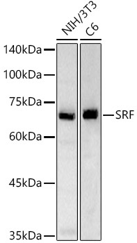
Western blot analysis of various lysates, using SRF Rabbit pAb (CAB7731) at 1:900 dilution. Secondary antibody: HRP-conjugated Goat anti-Rabbit IgG (H+L) (CABS014) at 1:10000 dilution. Lysates/proteins: 25μg per lane. Blocking buffer: 3% nonfat dry milk in TBST. Detection: ECL Basic Kit (AbGn00020). Exposure time: 60s.
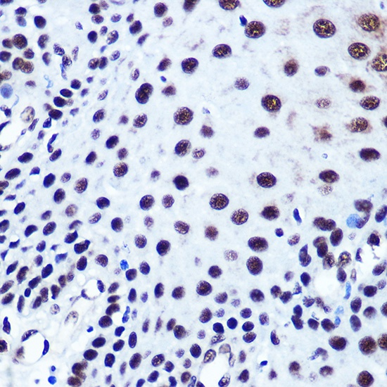
Immunohistochemistry analysis of paraffin-embedded Human esophageal using SRF Rabbit pAb (CAB7731) at dilution of 1:100 (40x lens). Microwave antigen retrieval performed with 0.01M PBS Buffer (pH 7.2) prior to IHC staining.
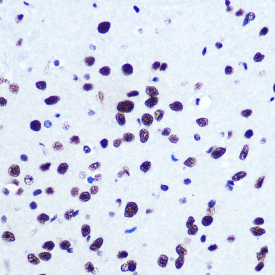
Immunohistochemistry analysis of paraffin-embedded Mouse brain using SRF Rabbit pAb (CAB7731) at dilution of 1:100 (40x lens). Microwave antigen retrieval performed with 0.01M PBS Buffer (pH 7.2) prior to IHC staining.