The SRPX Antibody (CAB1217) is a high-quality antibody developed for reliable detection and analysis of target proteins. Predicted to be an extracellular matrix structural constituent. Predicted to be involved in cell adhesion. Predicted to act upstream of or within several processes, including negative regulation of cell proliferation involved in contact inhibition; phagolysosome assembly; and positive regulation of extrinsic apoptotic signaling pathway in absence of ligand. Part of collagen-containing extracellular matrix.
This antibody is validated for use in WB, IHC-P, ELISA applications and has demonstrated reactivity against Human, Mouse samples.
Product Name:
SRPX Antibody
SKU:
CAB1217
Size:
100μL, 20μL
Reactivity:
Human, Mouse
Conjugate:
Unconjugated
Immunogen:
Recombinant protein (or fragment).This information is considered to be commercially sensitive.
Tested Applications:
WBIHC-PELISA
Recommended Dilution:
WB
1:500 - 1:2000
IHC-P
1:50 - 1:100
ELISA
Recommended starting concentration is 1 μg/mL. Please optimize the concentration based on your specific assay requirements.
Synonyms:
DRS, ETX1, SRPX1, HEL-S-83p, SRPX
Positive Sample:
Mouse heart
Cellular Localization:
Cell Surface.
Calculated MW:
52kDa
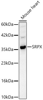
Observed MW:
35kDa
Predicted to be an extracellular matrix structural constituent. Predicted to be involved in cell adhesion. Predicted to act upstream of or within several processes, including negative regulation of cell proliferation involved in contact inhibition; phagolysosome assembly; and positive regulation of extrinsic apoptotic signaling pathway in absence of ligand. Part of collagen-containing extracellular matrix.
Purification Method
Affinity purification
Gene ID
8406
RRID
AB_2759056
Buffer Information
Store at -20℃. Avoid freeze / thaw cycles. Buffer: PBS containing 50% glycerol, preserved with proclin300 or sodium azide, pH 7.3.
Western blot analysis of lysates from Mouse heart, using SRPX Rabbit pAb (CAB1217) at 1:1000 dilution. Secondary antibody: HRP-conjugated Goat anti-Rabbit IgG (H+L) (AS014) at 1:10000 dilution. Lysates/proteins: 25μg per lane. Blocking buffer: 3% nonfat dry milk in TBST. Detection: ECL Basic Kit (AbGn00020). Exposure time: 30s.
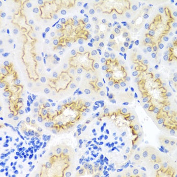
Immunohistochemistry analysis of paraffin-embedded Mouse kidney using SRPX Rabbit pAb (CAB1217) at dilution of 1:100 (40x lens). Microwave antigen retrieval performed with 0.01M PBS Buffer (pH 7.2) prior to IHC staining.