The SYT1 Antibody (CAB0992) is a high-quality antibody developed for reliable detection and analysis of target proteins. This gene encodes a member of the synaptotagmin protein family. The synaptotagmins are integral membrane proteins of synaptic vesicles that serve as calcium sensors in the process of vesicular trafficking and exocytosis. The encoded protein participates in triggering neurotransmitter release at the synapse in response to calcium binding. Mutations in this gene are associated with Baker-Gordon syndrome.
This antibody is validated for use in WB, IHC-P, ELISA, IF-P applications and has demonstrated reactivity against Human, Mouse, Rat samples.
Product Name:
SYT1 Antibody
SKU:
CAB0992
Size:
100μL, 20μL
Reactivity:
Human, Mouse, Rat
Conjugate:
Unconjugated
Immunogen:
Recombinant protein (or fragment).This information is considered to be commercially sensitive.
Tested Applications:
WBIHC-PELISAIF-P
Recommended Dilution:
WB
1:500 - 1:1000
IF-P
1:50 - 1:200
IHC-P
5000-20000
ELISA
Recommended starting concentration is 1 μg/mL. Please optimize the concentration based on your specific assay requirements.
This gene encodes a member of the synaptotagmin protein family. The synaptotagmins are integral membrane proteins of synaptic vesicles that serve as calcium sensors in the process of vesicular trafficking and exocytosis. The encoded protein participates in triggering neurotransmitter release at the synapse in response to calcium binding. Mutations in this gene are associated with Baker-Gordon syndrome.
Purification Method
Affinity purification
Gene ID
6857
RRID
AB_2757511
Buffer Information
Store at -20℃. Avoid freeze / thaw cycles. Buffer: PBS containing 50% glycerol, preserved with proclin300 or sodium azide, pH 7.3.
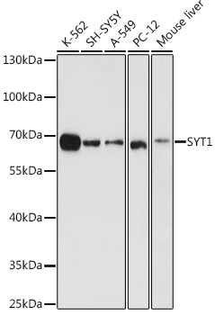
Western blot analysis of various lysates using SYT1 Rabbit pAb (CAB0992) at 1:1000 dilution. Secondary antibody: HRP-conjugated Goat anti-Rabbit IgG (H+L) (AS014) at 1:10000 dilution. Lysates/proteins: 25μg per lane. Blocking buffer: 3% nonfat dry milk in TBST. Detection: ECL Basic Kit (AbGn00020). Exposure time: 10s.
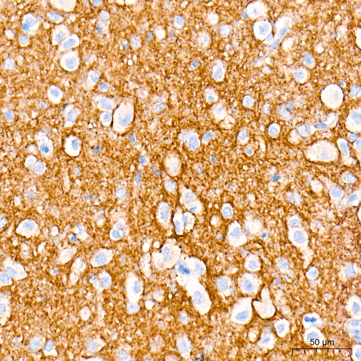
Immunohistochemistry analysis of paraffin-embedded Human brain tissue using SYT1 Rabbit pAb (CAB0992) at a dilution of 1:10000 (40x lens). High pressure antigen retrieval performed with 0.01M Tris-EDTA Buffer (pH 9.0) prior to IHC staining.
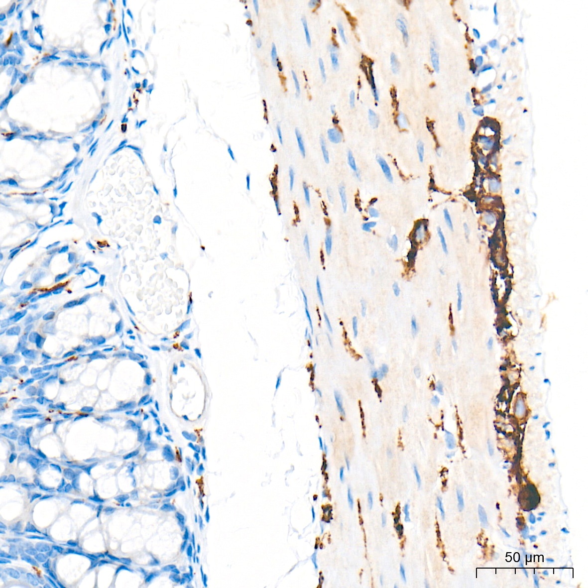
Immunohistochemistry analysis of paraffin-embedded Mouse colon tissue using SYT1 Rabbit pAb (CAB0992) at a dilution of 1:10000 (40x lens). High pressure antigen retrieval performed with 0.01M Tris-EDTA Buffer (pH 9.0) prior to IHC staining.
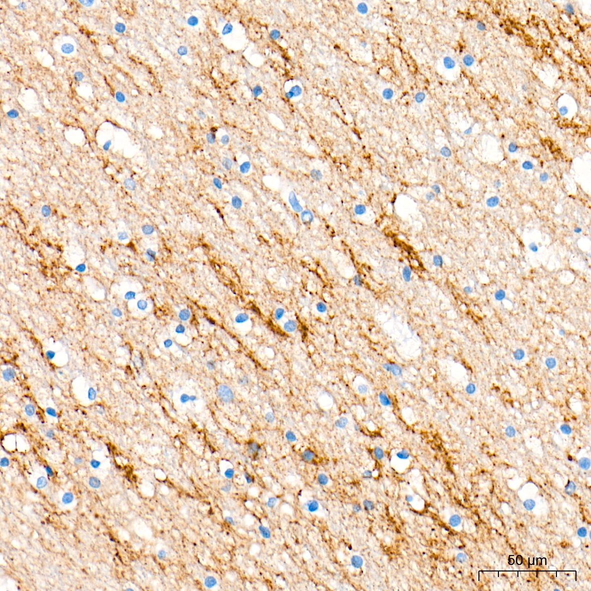
Immunohistochemistry analysis of paraffin-embedded Rat brain tissue using SYT1 Rabbit pAb (CAB0992) at a dilution of 1:10000 (40x lens). High pressure antigen retrieval performed with 0.01M Tris-EDTA Buffer (pH 9.0) prior to IHC staining.
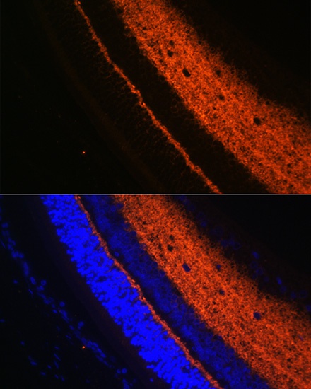
Immunofluorescence analysis of paraffin-embedded rat eye using SYT1 Rabbit pAb (CAB0992) at dilution of 1:100 (40x lens). Secondary antibody: Cy3-conjugated Goat anti-Rabbit IgG (H+L) (AS007) at 1:500 dilution. Blue: DAPI for nuclear staining.
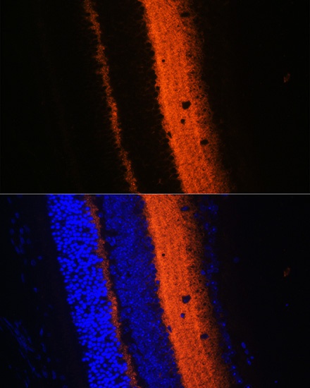
Immunofluorescence analysis of paraffin-embedded mouse eye using SYT1 Rabbit pAb (CAB0992) at dilution of 1:100 (40x lens). Secondary antibody: Cy3-conjugated Goat anti-Rabbit IgG (H+L) (AS007) at 1:500 dilution. Blue: DAPI for nuclear staining.


")


