The TAC3 Antibody (CAB6312) is a high-quality antibody developed for reliable detection and analysis of target proteins. This antibody, produced in rabbits, exhibits high reactivity with human samples and is suitable for use in Western blot applications.Tachykinin 3 is associated with functions such as regulation of the neuroendocrine system, pain perception, and inflammatory responses. By specifically binding to the tachykinin 3 protein, this antibody enables accurate detection and analysis in different cell types, making it an ideal choice for studies in neuroscience, endocrinology, and pain research.
This antibody is validated for use in WB, ELISA, IF-P applications and has demonstrated reactivity against Human, Mouse, Rat samples.
Product Name:
TAC3 Antibody
SKU:
CAB6312
Size:
20μL, 100μL
Reactivity:
Human, Mouse, Rat
Conjugate:
Unconjugated
Immunogen:
Recombinant protein (or fragment).This information is considered to be commercially sensitive.
Jurkat, Mouse spleen, Mouse brain, Rat kidney, Rat brain
Cellular Localization:
Secreted.
Calculated MW:
13kDa
Observed MW:
16kDa
This gene encodes a member of the tachykinin family of secreted neuropeptides. The encoded preproprotein is proteolytically processed to generate the mature peptide, which is primarily expressed in the central and peripheral nervous systems and functions as a neurotransmitter. This peptide is the ligand for the neurokinin-3 receptor. This protein is also expressed in the outer syncytiotrophoblast of the placenta and may be associated with pregnancy-induced hypertension and pre-eclampsia. Mutations in this gene are associated with normosmic hypogonadotropic hypogonadism. Alternative splicing results in multiple transcript variants, at least one of which encodes an isoform that is proteolytically processed.
Purification Method
Affinity purification
Gene ID
6866
RRID
AB_2766917
Buffer Information
Store at -20℃. Avoid freeze / thaw cycles. Buffer: PBS containing 50% glycerol, preserved with proclin300 or sodium azide, pH 7.3.
Western blot analysis of various lysates using TAC3 Rabbit pAb (CAB6312) at 1:1000 dilution._Secondary antibody: HRP-conjugated Goat anti-Rabbit IgG (H+L) (CABS014) at 1:10000 dilution._Lysates/proteins: 25μg per lane._Blocking buffer: 3% nonfat dry milk in TBST._Detection: ECL Enhanced Kit (AbGn00021)._Exposure time: 20s.
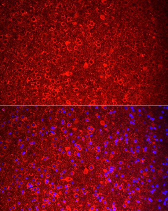
Immunofluorescence analysis of paraffin-embedded mouse brain using TAC3 Rabbit pAb (CAB6312) at dilution of 1:100 (40x lens). Secondary antibody: Cy3-conjugated Goat anti-Rabbit IgG (H+L) (CABS007) at 1:500 dilution. Blue: DAPI for nuclear staining.Perform microwave antigen retrieval with 0.01 M citrate buffer (pH 6.0) prior to IF staining.