The TAP1 Monoclonal Antibody (CAB22769) is a high-quality antibody developed for reliable detection and analysis of target proteins. This antibody, produced using hybridoma technology, specifically targets the TAP1 protein, which plays a key role in the transport of peptides for antigen presentation. With high specificity and sensitivity in detecting TAP1 in human samples, this antibody is ideal for use in techniques such as Western blotting and immunohistochemistry. By binding to TAP1, researchers can analyze the expression and function of this protein in various cell types and tissues, elucidating its role in immune responses and potential implications in diseases such as cancer and autoimmune disorders.
This antibody is validated for use in WB, ELISA applications and has demonstrated reactivity against Human, Mouse samples.
Product Name:
TAP1 Monoclonal Antibody
SKU:
CAB22769
Size:
20μL, 100μL
Reactivity:
Human, Mouse
Clone Number:
ARC59005
Conjugate:
Unconjugated
Immunogen:
Recombinant protein (or fragment).This information is considered to be commercially sensitive.
The membrane-associated protein encoded by this gene is a member of the superfamily of ATP-binding cassette (ABC) transporters. ABC proteins transport various molecules across extra- and intra-cellular membranes. ABC genes are divided into seven distinct subfamilies (ABC1, MDR/TAP, MRP, ALD, OABP, GCN20, White). This protein is a member of the MDR/TAP subfamily. Members of the MDR/TAP subfamily are involved in multidrug resistance. The protein encoded by this gene is involved in the pumping of degraded cytosolic peptides across the endoplasmic reticulum into the membrane-bound compartment where class I molecules assemble. Mutations in this gene may be associated with ankylosing spondylitis, insulin-dependent diabetes mellitus, and celiac disease. Two transcript variants encoding different isoforms have been found for this gene.
Purification Method
Affinity purification
Gene ID
6890
Buffer Information
Store at -20℃. Avoid freeze / thaw cycles. Buffer: PBS containing 50% glycerol and 0.05% BSA, preserved with proclin300 or sodium azide, pH 7.3.
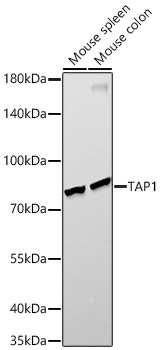
Western blot analysis of various lysates, using TAP1 Rabbit mAb (CAB22769) at 1:10000 dilution. Secondary antibody: HRP-conjugated Goat anti-Rabbit IgG (H+L) (CABS014) at 1:10000 dilution. Lysates/proteins: 25μg per lane. Blocking buffer: 3% nonfat dry milk in TBST. Detection: ECL Enhanced Kit (AbGn00021). Exposure time: 90s.
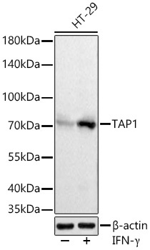
Western blot analysis of lysates from HT-29 cells using TAP1 Rabbit mAb (CAB22769) at 1:20000 dilution incubated overnight at 4℃. HT-29 cells were treated with IFNγ (50 ng/mL) for 8 hours. Secondary antibody: HRP-conjugated Goat anti-Rabbit IgG (H+L) (CABS014) at 1:10000 dilution. Lysates/proteins: 30 μg per lane. Blocking buffer: 3% nonfat dry milk in TBST. Detection: ECL Basic Kit (AbGn00020). Exposure time: 1s.
 at 1:10000 dilution. Secondary antibody: HRP Goat Anti-Rabbit IgG (H+L) at 1:10000 dilution. Lysates/proteins: 25μg per lane. Blocking buffer: 3% nonfat dry milk in TBST.")
 at 1:10000 dilution. Secondary antibody: HRP Goat Anti-Rabbit IgG (H+L) at 1:10000 dilution. Lysates/proteins: 25μg per lane. Blocking buffer: 3% nonfat dry milk in TBST.")


 at 1:10000 dilution. Lysates/proteins: 25ug per lane. Blocking buffer: 3% nonfat dry milk in TBST. Detection: ECL Basic Kit. Exposure time: 180s.")
 at 1:10000 dilution. Lysates/proteins: 25ug per lane. Blocking buffer: 3% nonfat dry milk in TBST. Detection: ECL Enhanced Kit. Exposure time: 45s.")