The TBK1/NAK Antibody (CAB2573) is a high-quality antibody developed for reliable detection and analysis of target proteins. This antibody, produced in rabbits, is highly specific to human samples and has been validated for use in Western blot applications. By binding to the TBK1 protein, this antibody enables precise detection and analysis in a variety of cell types, making it ideal for studies in immunology, cancer research, and beyond.TBK1, also known as TANK-binding kinase 1, is a crucial mediator of innate immune responses, playing a vital role in the activation of interferon-regulatory factors and the induction of type I interferons.
This antibody is validated for use in WB, IHC-P, ELISA applications and has demonstrated reactivity against Human, Mouse, Rat samples.
Product Name:
TBK1/NAK Antibody
SKU:
CAB2573
Size:
20μL, 100μL
Reactivity:
Human, Mouse, Rat
Conjugate:
Unconjugated
Immunogen:
Synthetic peptide. This information is considered to be commercially sensitive.
Recommended starting concentration is 1 μg/mL. Please optimize the concentration based on your specific assay requirements.
Synonyms:
NAK, T2K, IIAE8, FTDALS4, TBK1/NAK
Positive Sample:
HepG2, HeLa, Mouse lung, Rat thymus
Cellular Localization:
Cytoplasm.
Calculated MW:
84kDa
Observed MW:
84kDa
The NF-kappa-B (NFKB) complex of proteins is inhibited by I-kappa-B (IKB) proteins, which inactivate NFKB by trapping it in the cytoplasm. Phosphorylation of serine residues on the IKB proteins by IKB kinases marks them for destruction via the ubiquitination pathway, thereby allowing activation and nuclear translocation of the NFKB complex. The protein encoded by this gene is similar to IKB kinases and can mediate NFKB activation in response to certain growth factors. The protein is also an important kinase for antiviral innate immunity response.
Purification Method
Affinity purification
Gene ID
29110
RRID
AB_2764459
Buffer Information
Store at -20℃. Avoid freeze / thaw cycles. Buffer: PBS containing 50% glycerol, preserved with proclin300 or sodium azide, pH 7.3.
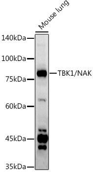
Western blot analysis of lysates from Mouse lung, using TBK1/NAK Rabbit pAb (CAB2573) at 1:1000 dilution. Secondary antibody: HRP-conjugated Goat anti-Rabbit IgG (H+L) (CABS014) at 1:10000 dilution. Lysates/proteins: 25μg per lane. Blocking buffer: 3% nonfat dry milk in TBST. Detection: ECL Basic Kit (AbGn00020). Exposure time: 90s.
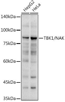
Western blot analysis of various lysates using TBK1/NAK Rabbit pAb (CAB2573) at 1:1000 dilution. Secondary antibody: HRP-conjugated Goat anti-Rabbit IgG (H+L) (CABS014) at 1:10000 dilution. Lysates/proteins: 25μg per lane. Blocking buffer: 3% nonfat dry milk in TBST. Detection: ECL Basic Kit (AbGn00020). Exposure time: 30s.
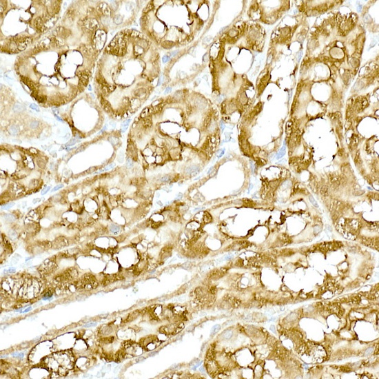
Immunohistochemistry analysis of paraffin-embedded Rat kidney using TBK1/NAK Rabbit pAb (CAB2573) at dilution of 1:200 (40x lens). High pressure antigen retrieval performed with 0.01M Citrate buffer (pH 6.0) prior to IHC staining.
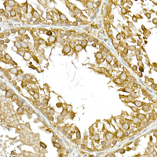
Immunohistochemistry analysis of paraffin-embedded Rat testis using TBK1/NAK Rabbit pAb (CAB2573) at dilution of 1:200 (40x lens). High pressure antigen retrieval performed with 0.01M Citrate buffer (pH 6.0) prior to IHC staining.