The TBRG4 Antibody (CAB15753) is a high-quality antibody developed for reliable detection and analysis of target proteins. This antibody, generated in rabbits, exhibits high reactivity with human samples and is validated for use in Western blot applications. By specifically binding to the TBRG4 protein, researchers can effectively detect and analyze its expression in various cell types, making it an indispensable resource for studies in cancer biology and cell signaling pathways.TBRG4, also known as TBRG4 Growth Factor 1, is involved in promoting cell division and survival, making it a potential target for cancer therapy and interventions targeting abnormal cell proliferation.
This antibody is validated for use in WB, IF/ICC, ELISA applications and has demonstrated reactivity against Human, Mouse, Rat samples.
Product Name:
TBRG4 Antibody
SKU:
CAB15753
Size:
20μL, 100μL
Reactivity:
Human, Mouse, Rat
Conjugate:
Unconjugated
Immunogen:
Recombinant protein (or fragment).This information is considered to be commercially sensitive.
Recommended starting concentration is 1 μg/mL. Please optimize the concentration based on your specific assay requirements.
Synonyms:
CPR2, FASTKD4, TBRG4
Positive Sample:
HepG2
Cellular Localization:
Mitochondrion.
Calculated MW:
71kDa
Observed MW:
68-71kDa
Enables RNA binding activity. Involved in mitochondrial mRNA processing and regulation of mitochondrial mRNA stability. Located in mitochondrial matrix.
Purification Method
Affinity purification
Gene ID
9238
RRID
AB_2763170
Buffer Information
Store at -20℃. Avoid freeze / thaw cycles. Buffer: PBS containing 50% glycerol, preserved with proclin300 or sodium azide, pH 7.3.
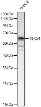
Western blot analysis of lysates from HepG2 cells, using TBRG4 Rabbit pAb (CAB15753) at 1:2000 dilution. Secondary antibody: HRP-conjugated Goat anti-Rabbit IgG (H+L) (CABS014) at 1:10000 dilution. Lysates/proteins: 25μg per lane. Blocking buffer: 3% nonfat dry milk in TBST. Detection: ECL Basic Kit (AbGn00020). Exposure time: 30s.
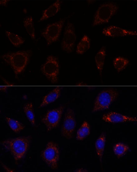
Immunofluorescence analysis of L929 cells using TBRG4 Rabbit pAb (CAB15753) at dilution of 1:100. Secondary antibody: Cy3-conjugated Goat anti-Rabbit IgG (H+L) (CABS007) at 1:500 dilution. Blue: DAPI for nuclear staining.