The TCF4 Antibody (CAB1141) is a high-quality antibody developed for reliable detection and analysis of target proteins. This antibody, generated in rabbits, exhibits high reactivity towards human samples and is validated for use in Western blot applications. By binding specifically to the TCF4 protein, this antibody allows for the detection and analysis of TCF4 expression in a variety of cell types, making it an ideal choice for studies in developmental biology and cancer research.TCF4, also known as transcription factor 7-like 2, is a key regulator of gene expression and plays a crucial role in controlling cell fate decisions.
This antibody is validated for use in WB, IF/ICC, ELISA applications and has demonstrated reactivity against Human, Mouse, Rat samples.
Product Name:
TCF4 Antibody
SKU:
CAB1141
Size:
20μL, 100μL
Reactivity:
Human, Mouse, Rat
Conjugate:
Unconjugated
Immunogen:
Recombinant protein (or fragment).This information is considered to be commercially sensitive.
This gene encodes transcription factor 4, a basic helix-loop-helix transcription factor. The encoded protein recognizes an Ephrussi-box ('E-box') binding site ('CANNTG') - a motif first identified in immunoglobulin enhancers. This gene is broadly expressed, and may play an important role in nervous system development. Defects in this gene are a cause of Pitt-Hopkins syndrome. In addition, an intronic CTG repeat normally numbering 10-37 repeat units can expand to >50 repeat units and cause Fuchs endothelial corneal dystrophy. Multiple alternatively spliced transcript variants that encode different proteins have been described.
Purification Method
Affinity purification
Gene ID
6925
RRID
AB_2758546
Buffer Information
Store at -20℃. Avoid freeze / thaw cycles. Buffer: PBS containing 50% glycerol, preserved with proclin300 or sodium azide, pH 7.3.
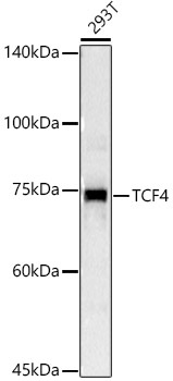
Western blot analysis of lysates from 293T cells, using TCF4 Rabbit pAb (CAB1141) at 1:1000 dilution. Secondary antibody: HRP-conjugated Goat anti-Rabbit IgG (H+L) (CABS014) at 1:10000 dilution. Lysates/proteins: 25μg per lane. Blocking buffer: 3% nonfat dry milk in TBST. Detection: ECL Basic Kit (AbGn00020). Exposure time: 3s.
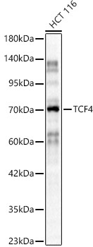
Western blot analysis of lysates from HCT 116 cells, using TCF4 Rabbit pAb (CAB1141) at 1:800 dilution. Secondary antibody: HRP-conjugated Goat anti-Rabbit IgG (H+L) (CABS014) at 1:10000 dilution. Lysates/proteins: 25μg per lane. Blocking buffer: 3% nonfat dry milk in TBST. Detection: ECL Basic Kit (AbGn00020). Exposure time: 30s.
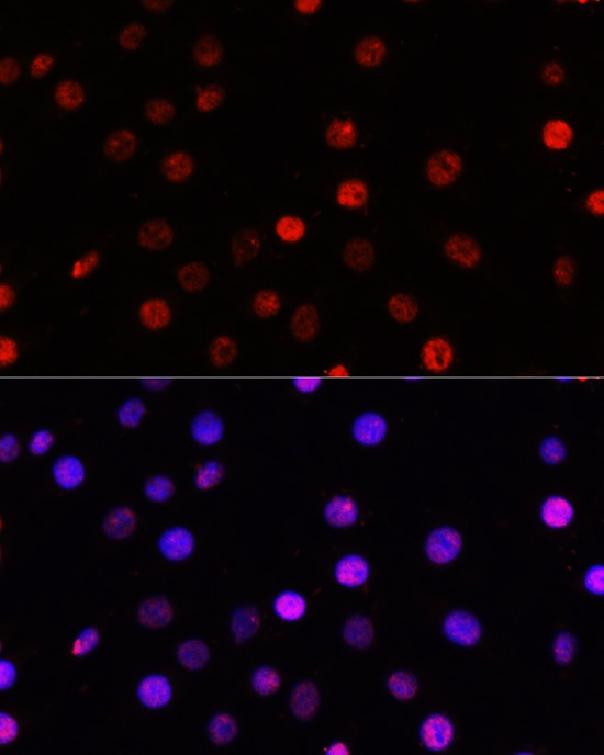
Immunofluorescence analysis of C6 cells using TCF4 Rabbit pAb (CAB1141) at dilution of 100 (40x lens). Secondary antibody: Cy3-conjugated Goat anti-Rabbit IgG (H+L) (CABS007) at 1:500 dilution. Blue: DAPI for nuclear staining.
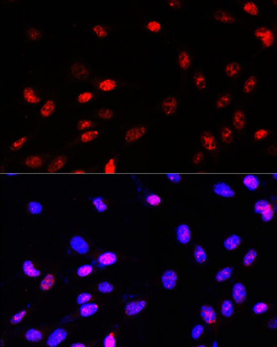
Immunofluorescence analysis of NIH-3T3 cells using TCF4 Rabbit pAb (CAB1141) at dilution of 100 (40x lens). Secondary antibody: Cy3-conjugated Goat anti-Rabbit IgG (H+L) (CABS007) at 1:500 dilution. Blue: DAPI for nuclear staining.


 at 1:10000 dilution. Lysates/proteins: 25ug per lane. Blocking buffer: 3% nonfat dry milk in TBST. Detection: ECL Basic Kit. Exposure time: 180s.")

