The TDG Antibody (CAB5756) is a high-quality antibody developed for reliable detection and analysis of target proteins. This rabbit-raised antibody is highly reactive with human samples and has been validated for use in Western blot applications. By binding specifically to the TDG protein, this antibody allows for the accurate detection and analysis of TDG expression levels in various cell types.TDG, or Thymine DNA Glycosylase, is essential for maintaining genomic integrity by removing aberrant or misincorporated thymine bases from DNA. Dysregulation of TDG activity has been implicated in various diseases, including cancer and neurodevelopmental disorders.
This antibody is validated for use in WB, ELISA applications and has demonstrated reactivity against Human, Mouse, Rat samples.
Product Name:
TDG Antibody
SKU:
CAB5756
Size:
20μL, 100μL
Reactivity:
Human, Mouse, Rat
Conjugate:
Unconjugated
Immunogen:
Recombinant protein (or fragment).This information is considered to be commercially sensitive.
Recommended starting concentration is 1 μg/mL. Please optimize the concentration based on your specific assay requirements.
Synonyms:
hTDG, TDG
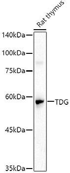
Positive Sample:
Rat thymus
Cellular Localization:
Nucleus.
Calculated MW:
46kDa
Observed MW:
50kDa
The protein encoded by this gene belongs to the TDG/mug DNA glycosylase family. Thymine-DNA glycosylase (TDG) removes thymine moieties from G/T mismatches by hydrolyzing the carbon-nitrogen bond between the sugar-phosphate backbone of DNA and the mispaired thymine. With lower activity, this enzyme also removes thymine from C/T and T/T mispairings. TDG can also remove uracil and 5-bromouracil from mispairings with guanine. This enzyme plays a central role in cellular defense against genetic mutation caused by the spontaneous deamination of 5-methylcytosine and cytosine. This gene may have a pseudogene in the p arm of chromosome 12.
Purification Method
Affinity purification
Gene ID
6996
RRID
AB_2766510
Buffer Information
Store at -20℃. Avoid freeze / thaw cycles. Buffer: PBS containing 50% glycerol, preserved with proclin300 or sodium azide, pH 7.3.
Western blot analysis of lysates from Rat thymus, using TDG Rabbit pAb (CAB5756) at 1:1000 dilution. Secondary antibody: HRP-conjugated Goat anti-Rabbit IgG (H+L) (CABS014) at 1:10000 dilution. Lysates/proteins: 25μg per lane. Blocking buffer: 3% nonfat dry milk in TBST. Detection: ECL Basic Kit (AbGn00020). Exposure time: 90s.