The TDGF1 Antibody (CAB1065) is a high-quality antibody developed for reliable detection and analysis of target proteins. This antibody, produced in rabbits, exhibits high reactivity with human samples and has been validated for Western blot applications. By specifically targeting the TDGF1 protein, researchers can accurately detect and analyze its expression in a variety of cell types, making it suitable for investigations in developmental biology, stem cell research, and cancer studies.
This antibody is validated for use in WB, IHC-P, ELISA applications and has demonstrated reactivity against Human, Mouse, Rat samples.
Product Name:
TDGF1 Antibody
SKU:
CAB1065
Size:
20μL, 100μL
Reactivity:
Human, Mouse, Rat
Conjugate:
Unconjugated
Immunogen:
Recombinant protein (or fragment).This information is considered to be commercially sensitive.
Recommended starting concentration is 1 μg/mL. Please optimize the concentration based on your specific assay requirements.
Synonyms:
CR, CR-1, CRGF, CRIPTO, TDGF1
Positive Sample:
NCCIT
Cellular Localization:
Cell Membrane, Gpi-Anchor, Lipid-Anchor.
Calculated MW:
21kDa
Observed MW:
18kDa/20kDa
This gene encodes an epidermal growth factor-related protein that contains a cripto, FRL-1, and cryptic domain. The encoded protein is an extracellular, membrane-bound signaling protein that plays an essential role in embryonic development and tumor growth. Mutations in this gene are associated with forebrain defects. Pseudogenes of this gene are found on chromosomes 2, 3, 6, 8, 19 and X. Alternate splicing results in multiple transcript variants.
Purification Method
Affinity purification
Gene ID
6997
RRID
AB_2758160
Buffer Information
Store at -20℃. Avoid freeze / thaw cycles. Buffer: PBS containing 50% glycerol, preserved with proclin300 or sodium azide, pH 7.3.
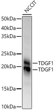
Western blot analysis of lysates from NCCIT cells, using TDGF1 Rabbit pAb (CAB1065) at 1:900 dilution. Secondary antibody: HRP-conjugated Goat anti-Rabbit IgG (H+L) (CABS014) at 1:10000 dilution. Lysates/proteins: 25μg per lane. Blocking buffer: 3% nonfat dry milk in TBST. Detection: ECL Basic Kit (AbGn00020). Exposure time: 60s.
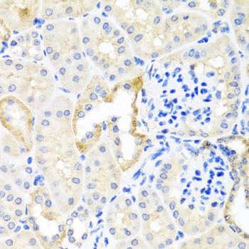
Immunohistochemistry analysis of paraffin-embedded Mouse kidney using TDGF1 Rabbit pAb (CAB1065) at dilution of 1:100 (40x lens). Microwave antigen retrieval performed with 0.01M PBS Buffer (pH 7.2) prior to IHC staining.
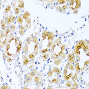
Immunohistochemistry analysis of paraffin-embedded Human stomach using TDGF1 Rabbit pAb (CAB1065) at dilution of 1:100 (40x lens). Microwave antigen retrieval performed with 0.01M PBS Buffer (pH 7.2) prior to IHC staining.