The TDP1 Antibody (CAB4849) is a high-quality antibody developed for reliable detection and analysis of target proteins. This antibody, raised in rabbits, has high reactivity with human samples and has been validated for use in Western blot applications. By binding to the TDP1 protein, this antibody enables accurate detection and analysis in a variety of cell types, making it suitable for research in fields such as cancer biology, genomics, and personalized medicine.TDP1, also known as tyrosyl-DNA phosphodiesterase 1, plays a crucial role in repairing DNA damage caused by various factors such as environmental toxins, radiation, and reactive oxygen species.
This antibody is validated for use in WB, IF/ICC, ELISA applications and has demonstrated reactivity against Human, Rat samples.
Product Name:
TDP1 Antibody
SKU:
CAB4849
Size:
20μL, 100μL
Reactivity:
Human, Rat
Conjugate:
Unconjugated
Immunogen:
Recombinant protein (or fragment).This information is considered to be commercially sensitive.
Recommended starting concentration is 1 μg/mL. Please optimize the concentration based on your specific assay requirements.
Synonyms:
TDP1
Positive Sample:
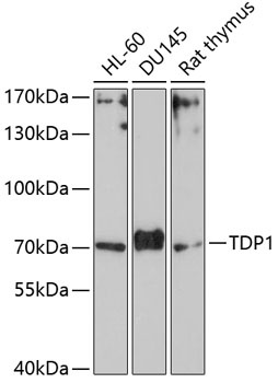
HL-60, DU145, Rat thymus
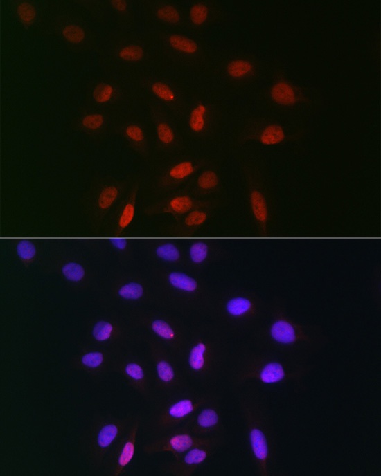
Cellular Localization:
Cytoplasm, Nucleus.
Calculated MW:
68kDa
Observed MW:
70kDa
The protein encoded by this gene is involved in repairing stalled topoisomerase I-DNA complexes by catalyzing the hydrolysis of the phosphodiester bond between the tyrosine residue of topoisomerase I and the 3-prime phosphate of DNA. This protein may also remove glycolate from single-stranded DNA containing 3-prime phosphoglycolate, suggesting a role in repair of free-radical mediated DNA double-strand breaks. This gene is a member of the phospholipase D family and contains two PLD phosphodiesterase domains. Mutations in this gene are associated with the disease spinocerebellar ataxia with axonal neuropathy (SCAN1).
Purification Method
Affinity purification
Gene ID
55775
RRID
AB_2765903
Buffer Information
Store at -20℃. Avoid freeze / thaw cycles. Buffer: PBS containing 50% glycerol, preserved with proclin300 or sodium azide, pH 7.3.
Western blot analysis of various lysates using TDP1 Rabbit pAb (CAB4849) at 1:1000 dilution. Secondary antibody: HRP-conjugated Goat anti-Rabbit IgG (H+L) (CABS014) at 1:10000 dilution. Lysates/proteins: 25μg per lane. Blocking buffer: 3% nonfat dry milk in TBST. Detection: ECL Basic Kit (AbGn00020). Exposure time: 90s.
Immunofluorescence analysis of U-2 OS cells using TDP1 Rabbit pAb (CAB4849) at dilution of 1:100 (40x lens). Secondary antibody: Cy3-conjugated Goat anti-Rabbit IgG (H+L) (CABS007) at 1:500 dilution. Blue: DAPI for nuclear staining.