The TET3 Monoclonal Antibody (CAB21930) is a high-quality antibody developed for reliable detection and analysis of target proteins. This antibody, produced through hybridoma technology, has high specificity and sensitivity for detecting TET3 in various sample types, including human tissues. Validated for use in techniques such as immunofluorescence and immunoprecipitation, this antibody enables precise localization and quantification of TET3 in cells and tissues.TET3, a member of the TET family of proteins, plays a critical role in the dynamic regulation of DNA methylation, which is essential for processes such as embryonic development and maintenance of pluripotency.
This antibody is validated for use in WB, ELISA applications and has demonstrated reactivity against Mouse samples.
Product Name:
TET3 Monoclonal Antibody
SKU:
CAB21930
Size:
20μL, 100μL
Reactivity:
Mouse
Clone Number:
ARC52663
Conjugate:
Unconjugated
Immunogen:
Recombinant protein (or fragment).This information is considered to be commercially sensitive.
Recommended starting concentration is 1 μg/mL. Please optimize the concentration based on your specific assay requirements.
Synonyms:
B430006D22Rik, D230004J03Rik, TET3
Positive Sample:
Neuro-2a, 293T transfected with TET3
Cellular Localization:
Cytoplasm, Female Pronucleus, Male Pronucleus, Nucleoplasm, Nucleus.
Calculated MW:
195kDa
Observed MW:
230kDa
Enables methylcytosine dioxygenase activity. Involved in DNA demethylation of male pronucleus. Acts upstream of or within DNA demethylation. Located in cytoplasm; female pronucleus; and male pronucleus. Is expressed in several structures, including central nervous system; early conceptus; future brain; and genitourinary system. Orthologous to human TET3 (tet methylcytosine dioxygenase 3).
Purification Method
Affinity purification
Gene ID
194388
Buffer Information
Store at -20℃. Avoid freeze / thaw cycles. Buffer: PBS containing 50% glycerol and 0.05% BSA, preserved with proclin300 or sodium azide, pH 7.3.
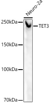
Western blot analysis of lysates from Neuro-2a cells, using TET3 Rabbit mAb (CAB21930) at 1:20000 dilution.Secondary antibody: HRP-conjugated Goat anti-Rabbit IgG (H+L) (CABS014) at 1:10000 dilution.Lysates/proteins: 25μg per lane.Blocking buffer: 3% nonfat dry milk in TBST.Detection: ECL Basic Kit (AbGn00020).Exposure time: 90s.
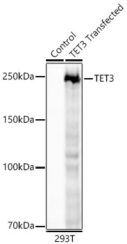
Western blot analysis of lysates from wild type (WT) and 293T cells transfected with TET3 using TET3 Rabbit mAb (CAB21930) at 1:20000 dilution incubated overnight at 4℃. Secondary antibody: HRP-conjugated Goat anti-Rabbit IgG (H+L) (CABS014) at 1:10000 dilution. Lysates/proteins: 25 μg per lane. Blocking buffer: 3% nonfat dry milk in TBST. Detection: ECL Basic Kit (AbGn00020) Exposure time: 30 s.
 at 1:20000 dilution. Secondary antibody: HRP Goat Anti-Rabbit IgG (H+L) at 1:10000 dilution. Lysates/proteins: 25μg per lane. Blocking buffer: 3% nonfat dry milk in TBST.")
 at 1:20000 dilution. Secondary antibody: HRP Goat Anti-Rabbit IgG (H+L) at 1:10000 dilution. Lysates/proteins: 25μg per lane. Blocking buffer: 3% nonfat dry milk in TBST.")


