The TFE3 Monoclonal Antibody (CAB0548) is a high-quality antibody developed for reliable detection and analysis of target proteins. This antibody, generated in rabbits, is highly specific to human samples and has been extensively validated for its use in immunofluorescence and immunohistochemistry applications.TFE3, a member of the microphthalmia (MiT) family of transcription factors, plays a crucial role in the regulation of gene expression involved in cell growth and development. Its dysregulation has been linked to various diseases, including cancer, metabolic disorders, and neurodegenerative diseases.
This antibody is validated for use in WB, IHC-P, ELISA applications and has demonstrated reactivity against Human, Mouse, Rat samples.
Product Name:
TFE3 Monoclonal Antibody
SKU:
CAB0548
Size:
20μL, 100μL
Reactivity:
Human, Mouse, Rat
Clone Number:
ARC1829
Conjugate:
Unconjugated
Immunogen:
Recombinant protein (or fragment).This information is considered to be commercially sensitive.
Recommended starting concentration is 1 μg/mL. Please optimize the concentration based on your specific assay requirements.
Synonyms:
TFEA, RCCP2, RCCX1, MRXSPF, bHLHe33, TFE3
Positive Sample:
NIH/3T3, C6, HeLa
Cellular Localization:
Nucleus.
Calculated MW:
62kDa
Observed MW:
70kDa
This gene encodes a basic helix-loop-helix domain-containing transcription factor that binds MUE3-type E-box sequences in the promoter of genes. The encoded protein promotes the expression of genes downstream of transforming growth factor beta (TGF-beta) signaling. This gene may be involved in chromosomal translocations in renal cell carcinomas and other cancers, resulting in the production of fusion proteins. Translocation partners include PRCC (papillary renal cell carcinoma), NONO (non-POU domain containing, octamer-binding), and ASPSCR1 (alveolar soft part sarcoma chromosome region, candidate 1), among other genes. Alternative splicing results in multiple transcript variants.
Purification Method
Affinity purification
Gene ID
7030
RRID
AB_2861464
Buffer Information
Store at -20℃. Avoid freeze / thaw cycles. Buffer: PBS containing 50% glycerol and 0.05% BSA, preserved with proclin300 or sodium azide, pH 7.3.
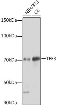
Western blot analysis of various lysates using TFE3 Rabbit mAb (CAB0548) at 1:1000 dilution. Secondary antibody: HRP-conjugated Goat anti-Rabbit IgG (H+L) (CABS014) at 1:10000 dilution. Lysates/proteins: 25μg per lane. Blocking buffer: 3% nonfat dry milk in TBST. Detection: ECL Basic Kit (AbGn00020). Exposure time: 30s.
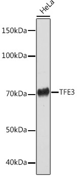
Western blot analysis of lysates from HeLa cells, using TFE3 Rabbit mAb (CAB0548) at 1:1000 dilution. Secondary antibody: HRP-conjugated Goat anti-Rabbit IgG (H+L) (CABS014) at 1:10000 dilution. Lysates/proteins: 25μg per lane. Blocking buffer: 3% nonfat dry milk in TBST. Detection: ECL Basic Kit (AbGn00020). Exposure time: 90s.