The TGM3 Polyclonal Antibody (CAB24495) is a high-quality antibody developed for reliable detection and analysis of target proteins. This antibody, raised in rabbits, is highly specific to human TGM3 and is validated for use in Western blot applications. By specifically binding to the TGM3 protein, researchers can accurately detect and analyze TGM3 levels in different cell types.TGM3 is a key enzyme involved in various biological processes, including skin barrier function, epidermal differentiation, and wound healing.
This antibody is validated for use in WB, IF/ICC, ELISA applications and has demonstrated reactivity against Human, Mouse samples.
Product Name:
TGM3 Polyclonal Antibody
SKU:
CAB24495
Size:
20μL, 100μL
Reactivity:
Human, Mouse
Conjugate:
Unconjugated
Immunogen:
Recombinant protein (or fragment).This information is considered to be commercially sensitive.
Recommended starting concentration is 1 μg/mL. Please optimize the concentration based on your specific assay requirements.
Synonyms:
TGM3, TGE, UHS2, transglutaminase 3
Positive Sample:
293T, 293T-TGM35-His(C)
Cellular Localization:
Cytoplasm, Extracellular Exosome.
Calculated MW:
76kDa
Observed MW:
70kDa
Transglutaminases are enzymes that catalyze the crosslinking of proteins by epsilon-gamma glutamyl lysine isopeptide bonds. While the primary structure of transglutaminases is not conserved, they all have the same amino acid sequence at their active sites and their activity is calcium-dependent. The protein encoded by this gene consists of two polypeptide chains activated from a single precursor protein by proteolysis. The encoded protein is involved the later stages of cell envelope formation in the epidermis and hair follicle.
Purification Method
Affinity purification
Gene ID
7053
Buffer Information
Store at -20℃. Avoid freeze / thaw cycles. Buffer: PBS containing 50% glycerol, preserved with proclin300 or sodium azide, pH 7.3.
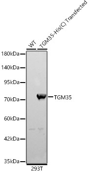
Western blot analysis of lysates from wild type (WT) and 293T cells transfected with TGM3 using TGM3 Rabbit pAb (CAB24495) at 1:2000 dilution. Secondary antibody: HRP-conjugated Goat anti-Rabbit IgG (H+L) (CABS014) at 1:10000 dilution. Lysates / proteins: 25 μg per lane. Blocking buffer: 3 % nonfat dry milk in TBST. Detection: ECL Basic Kit (AbGn00020). Exposure time: 10s.
 and 293T cells transfected with TGM3 using TGM3 Rabbit pAb (CAB24495) at 1:2000 dilution. Secondary antibody:HRP Goat Anti-Rabbit IgG (H+L) at 1:10000 dilution. Lysates / proteins: 25 μg per lane. Blocking buffer: 3 % nonfat dry milk in TBST. Detection:ECL Basic Kit (RM00020). Exposuretime: 10s.")
 and 293T cells transfected with TGM3 using TGM3 Rabbit pAb (CAB24495) at 1:2000 dilution. Secondary antibody:HRP Goat Anti-Rabbit IgG (H+L) at 1:10000 dilution. Lysates / proteins: 25 μg per lane. Blocking buffer: 3 % nonfat dry milk in TBST. Detection:ECL Basic Kit (RM00020). Exposuretime: 10s.")


")
 at 1:10000 dilution. Lysates/proteins: 25ug per lane. Blocking buffer: 3% nonfat dry milk in TBST. Detection: ECL Basic Kit. Exposure time: 1s.")