The THOC7 Antibody (CAB13700) is a high-quality antibody developed for reliable detection and analysis of target proteins. This antibody, generated in rabbits, is highly specific to human samples and has been validated for use in Western blot applications.THOC7 is essential for mRNA export and its dysregulation has been linked to various diseases including cancer and neurodegenerative disorders.
This antibody is validated for use in WB, ELISA applications and has demonstrated reactivity against Human, Mouse, Rat samples.
Product Name:
THOC7 Antibody
SKU:
CAB13700
Size:
20μL, 100μL
Reactivity:
Human, Mouse, Rat
Conjugate:
Unconjugated
Immunogen:
Recombinant protein (or fragment).This information is considered to be commercially sensitive.
Recommended starting concentration is 1 μg/mL. Please optimize the concentration based on your specific assay requirements.
Synonyms:
fSAP24, hTREX30, NIF3L1BP1, THOC7
Positive Sample:
HT-29, A-431, 293T, Jurkat, Mouse kidney, Rat brain
Cellular Localization:
Cytoplasm, Nucleus, Nucleus Speckle.
Calculated MW:
24kDa
Observed MW:
24kDa
Predicted to enable RNA binding activity. Involved in mRNA export from nucleus and viral mRNA export from host cell nucleus. Located in cytosol and nuclear speck. Part of THO complex part of transcription export complex. Colocalizes with chromosome, telomeric region.
Purification Method
Affinity purification
Gene ID
80145
RRID
AB_2760561
Buffer Information
Store at -20℃. Avoid freeze / thaw cycles. Buffer: PBS with 0.01% thimerosal,50% glycerol,pH7.3.
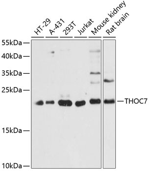
Western blot analysis of various lysates using THOC7 Rabbit pAb (CAB13700) at 1:3000 dilution. Secondary antibody: HRP-conjugated Goat anti-Rabbit IgG (H+L) (CABS014) at 1:10000 dilution. Lysates/proteins: 25μg per lane. Blocking buffer: 3% nonfat dry milk in TBST. Detection: ECL Basic Kit (AbGn00020). Exposure time: 60s.