The DTYMK Antibody (CAB6370) is a high-quality antibody developed for reliable detection and analysis of target proteins. This antibody, generated in rabbits, exhibits high reactivity with human samples and is validated for use in Western blot applications. By targeting the thymidylate kinase protein, this antibody allows for precise detection and analysis in a variety of cell types, making it an excellent choice for studies in molecular biology and cancer research.
This antibody is validated for use in WB, IF/ICC, ELISA applications and has demonstrated reactivity against Human, Mouse, Rat samples.
Product Name:
DTYMK Antibody
SKU:
CAB6370
Size:
20μL, 100μL
Reactivity:
Human, Mouse, Rat
Conjugate:
Unconjugated
Immunogen:
Recombinant protein (or fragment).This information is considered to be commercially sensitive.
Recommended starting concentration is 1 μg/mL. Please optimize the concentration based on your specific assay requirements.
Synonyms:
CDC8, TMPK, TYMK, CONPM, PP3731, DTYMK
Positive Sample:
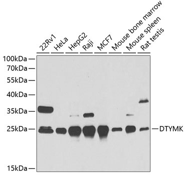
22Rv1, HeLa, HepG2, Raji, MCF7, Mouse bone marrow, Mouse spleen, Rat testis
Cellular Localization:
Cytoplasm, Cytosol, Mitochondrion, Nucleus.
Calculated MW:
24kDa
Observed MW:
24kDa
Enables thymidylate kinase activity. Predicted to be involved in dTDP biosynthetic process; dTTP biosynthetic process; and dUDP biosynthetic process. Predicted to act upstream of or within cellular response to growth factor stimulus and nucleotide biosynthetic process. Predicted to be located in mitochondrial intermembrane space and mitochondrial matrix. Predicted to be active in cytosol; mitochondrion; and nucleus.
Purification Method
Affinity purification
Gene ID
1841
RRID
AB_2766972
Buffer Information
Store at -20℃. Avoid freeze / thaw cycles. Buffer: PBS containing 50% glycerol, preserved with proclin300 or sodium azide, pH 7.3.
Western blot analysis of various lysates using DTYMK Rabbit pAb (CAB6370) at 1:1000 dilution. Secondary antibody: HRP-conjugated Goat anti-Rabbit IgG (H+L) (CABS014) at 1:10000 dilution. Lysates/proteins: 25μg per lane. Blocking buffer: 3% nonfat dry milk in TBST. Detection: ECL Basic Kit (AbGn00020). Exposure time: 30s.
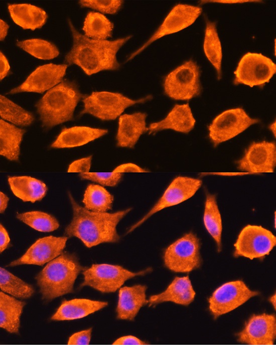
Immunofluorescence analysis of L929 cells using DTYMK Rabbit pAb (CAB6370) at dilution of 1:100 (40x lens). Secondary antibody: Cy3-conjugated Goat anti-Rabbit IgG (H+L) (CABS007) at 1:500 dilution. Blue: DAPI for nuclear staining.