The TIMM8A Antibody (CAB9811) is a high-quality antibody developed for reliable detection and analysis of target proteins. This antibody, produced in rabbits, is highly specific to human samples and has been validated for use in Western blot applications. By binding to the TIMM8A protein, this antibody enables researchers to detect and analyze TIMM8A in a variety of cell types, making it ideal for studies in mitochondrial biology and related research areas.TIMM8A is a key component of the TIMM complex, responsible for importing nuclear-encoded proteins into the mitochondria.
This antibody is validated for use in WB, IF/ICC, ELISA applications and has demonstrated reactivity against Human, Mouse, Rat samples.
Product Name:
TIMM8A Antibody
SKU:
CAB9811
Size:
20μL, 100μL
Reactivity:
Human, Mouse, Rat
Conjugate:
Unconjugated
Immunogen:
Recombinant protein (or fragment).This information is considered to be commercially sensitive.
This translocase is involved in the import and insertion of hydrophobic membrane proteins from the cytoplasm into the mitochondrial inner membrane. The gene is mutated in Mohr-Tranebjaerg syndrome/Deafness Dystonia Syndrome (MTS/DDS) and it is postulated that MTS/DDS is a mitochondrial disease caused by a defective mitochondrial protein import system. Defects in this gene also cause Jensen syndrome; an X-linked disease with opticoacoustic nerve atrophy and muscle weakness. This protein, along with TIMM13, forms a 70 kDa heterohexamer. Alternative splicing results in multiple transcript variants encoding distinct isoforms.
Purification Method
Affinity purification
Gene ID
1678
RRID
AB_2772614
Buffer Information
Store at -20℃. Avoid freeze / thaw cycles. Buffer: PBS containing 50% glycerol, preserved with proclin300 or sodium azide, pH 7.3.
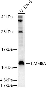
Western blot analysis of lysates from U-87MG cells, using TIMM8A Rabbit pAb (CAB9811) at 1:600 dilution. Secondary antibody: HRP-conjugated Goat anti-Rabbit IgG (H+L) (CABS014) at 1:10000 dilution. Lysates/proteins: 25μg per lane. Blocking buffer: 3% nonfat dry milk in TBST. Detection: ECL Enhanced Kit (AbGn00021). Exposure time: 90s.
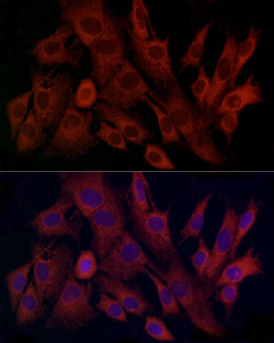
Immunofluorescence analysis of C6 cells using TIMM8A Rabbit pAb (CAB9811) at dilution of 1:100 (40x lens). Secondary antibody: Cy3-conjugated Goat anti-Rabbit IgG (H+L) (CABS007) at 1:500 dilution. Blue: DAPI for nuclear staining.