The TLR3 Antibody (CAB11778) is a high-quality antibody developed for reliable detection and analysis of target proteins. This antibody, produced in rabbits, demonstrates high reactivity with human samples and is validated for Western blot applications. By specifically binding to the TLR3 protein, this antibody enables precise detection and analysis in a variety of cell types, making it ideal for studies in immunology and infectious disease research.TLR3 is a critical component of the immune system, recognizing double-stranded RNA from viruses and triggering a cascade of immune responses to combat infection.
This antibody is validated for use in WB, IHC-P, IF/ICC, ELISA applications and has demonstrated reactivity against Human, Mouse, Rat samples.
Product Name:
TLR3 Antibody
SKU:
CAB11778
Size:
20μL, 100μL
Reactivity:
Human, Mouse, Rat
Conjugate:
Unconjugated
Immunogen:
Recombinant protein (or fragment).This information is considered to be commercially sensitive.
Recommended starting concentration is 1 μg/mL. Please optimize the concentration based on your specific assay requirements.
Synonyms:
CD283, IIAE2, IMD83, TLR3
Positive Sample:
HeLa, Rat liver, Mouse lung
Cellular Localization:
Early Endosome, Endoplasmic Reticulum Membrane, Endosome Membrane, Single-Pass Type I Membrane Protein.
Calculated MW:
104kDa
Observed MW:
104kDa
The protein encoded by this gene is a member of the Toll-like receptor (TLR) family which plays a fundamental role in pathogen recognition and activation of innate immunity. TLRs are highly conserved from Drosophila to humans and share structural and functional similarities. They recognize pathogen-associated molecular patterns (PAMPs) that are expressed on infectious agents, and mediate the production of cytokines necessary for the development of effective immunity. The various TLRs exhibit different patterns of expression. This receptor is most abundantly expressed in placenta and pancreas, and is restricted to the dendritic subpopulation of the leukocytes. It recognizes dsRNA associated with viral infection, and induces the activation of NF-kappaB and the production of type I interferons. It thus plays a role in host defense against multiple viruses.
Purification Method
Affinity purification
Gene ID
7098
RRID
AB_2758748
Buffer Information
Store at -20℃. Avoid freeze / thaw cycles. Buffer: PBS containing 50% glycerol, preserved with proclin300 or sodium azide, pH 7.3.
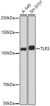
Western blot analysis of various lysates using TLR3 Rabbit pAb (CAB11778) at 1:1000 dilution. Secondary antibody: HRP-conjugated Goat anti-Rabbit IgG (H+L) (CABS014) at 1:10000 dilution. Lysates/proteins: 25μg per lane. Blocking buffer: 3% nonfat dry milk in TBST. Detection: ECL Basic Kit (AbGn00020). Exposure time: 90s.
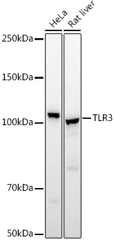
Western blot analysis of various lysates using TLR3 Rabbit pAb (CAB11778) at 1:500 dilution. Secondary antibody: HRP-conjugated Goat anti-Rabbit IgG (H+L) (CABS014) at 1:10000 dilution. Lysates/proteins: 25μg per lane. Blocking buffer: 3% nonfat dry milk in TBST. Detection: ECL Basic Kit (AbGn00020). Exposure time: 10s.
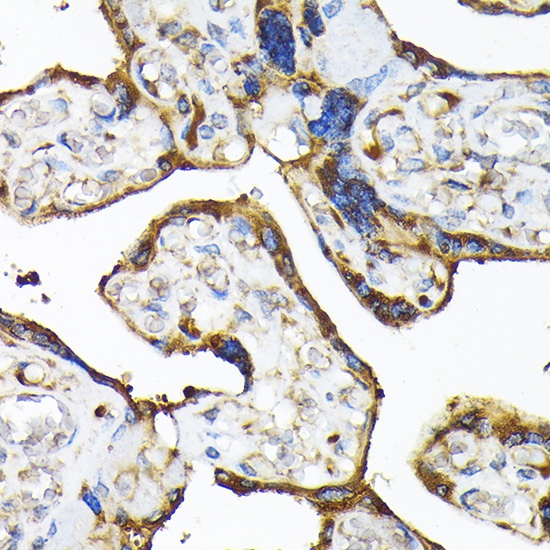
Immunohistochemistry analysis of paraffin-embedded Human placenta using TLR3 Rabbit pAb (CAB11778) at dilution of 1:100 (40x lens). Microwave antigen retrieval performed with 0.01M Tris/EDTA Buffer (pH 9.0) prior to IHC staining.
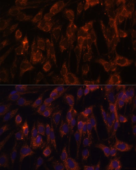
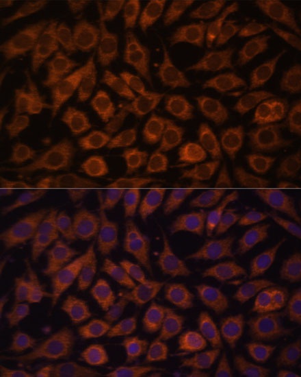
Immunofluorescence analysis of C6 cells using TLR3 Rabbit pAb (CAB11778) at dilution of 1:100. Secondary antibody: Cy3-conjugated Goat anti-Rabbit IgG (H+L) (CABS007) at 1:500 dilution. Blue: DAPI for nuclear staining.
Immunofluorescence analysis of L929 cells using TLR3 Rabbit pAb (CAB11778) at dilution of 1:100. Secondary antibody: Cy3-conjugated Goat anti-Rabbit IgG (H+L) (CABS007) at 1:500 dilution. Blue: DAPI for nuclear staining.


")


