The TMED2 Antibody (CAB17622) is a high-quality antibody developed for reliable detection and analysis of target proteins. This rabbit polyclonal antibody is highly specific to human samples and has been validated for use in Western blot applications. By binding to the TMED2 protein, this antibody enables the detection and analysis of TMED2 in a variety of cell types, making it an ideal choice for studies in cell biology and molecular biology.
This antibody is validated for use in WB, IF/ICC, ELISA applications and has demonstrated reactivity against Human, Mouse, Rat samples.
Product Name:
TMED2 Antibody
SKU:
CAB17622
Size:
20μL, 100μL
Reactivity:
Human, Mouse, Rat
Conjugate:
Unconjugated
Immunogen:
Recombinant protein (or fragment).This information is considered to be commercially sensitive.
Recommended starting concentration is 1 μg/mL. Please optimize the concentration based on your specific assay requirements.
Synonyms:
p24, P24A, RNP24, p24b1, p24beta1, TMED2
Positive Sample:
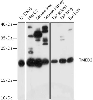
U-87MG, HepG2, Mouse liver, Mouse kidney, Rat spleen, Rat lung, Rat liver
Cellular Localization:
Copi-Coated Vesicle, Copii-Coated Er To Golgi Transport Vesicle, Endoplasmic Reticulum, Endoplasmic Reticulum-Golgi Intermediate Compartment, Golgi Apparatus, Transport Vesicle, Zymogen Granule Membrane.
Calculated MW:
23kDa
Observed MW:
23kDa
Involved in several processes, including Golgi organization; negative regulation of GTPase activity; and protein localization to plasma membrane. Located in Golgi apparatus; endoplasmic reticulum; and endoplasmic reticulum-Golgi intermediate compartment.
Purification Method
Affinity purification
Gene ID
10959
RRID
AB_2772632
Buffer Information
Store at -20℃. Avoid freeze / thaw cycles. Buffer: PBS with 0.01% thimerosal,50% glycerol,pH7.3.
Western blot analysis of various lysates using TMED2 Rabbit pAb (CAB17622) at 1:1000 dilution. Secondary antibody: HRP-conjugated Goat anti-Rabbit IgG (H+L) (CABS014) at 1:10000 dilution. Lysates/proteins: 25μg per lane. Blocking buffer: 3% nonfat dry milk in TBST. Detection: ECL Basic Kit (AbGn00020). Exposure time: 3min.
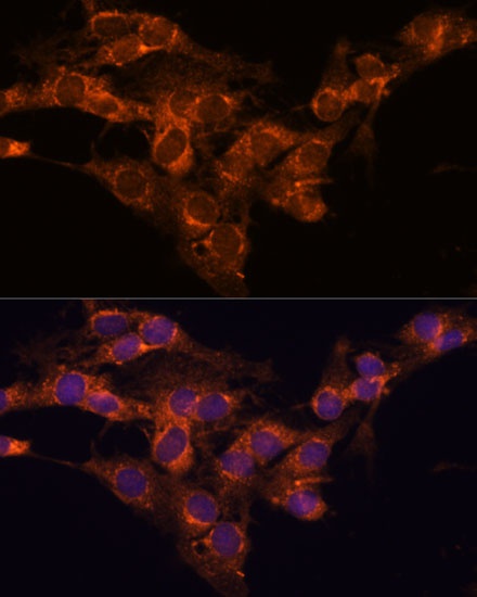
Immunofluorescence analysis of C6 cells using TMED2 Rabbit pAb (CAB17622) at dilution of 1:100. Secondary antibody: Cy3-conjugated Goat anti-Rabbit IgG (H+L) (CABS007) at 1:500 dilution. Blue: DAPI for nuclear staining.