The TMX1 Antibody (CAB14936) is a high-quality antibody developed for reliable detection and analysis of target proteins. This antibody, generated in rabbits, is highly specific to human samples and has been validated for use in Western blot applications. By binding to the TMX1 protein, this antibody enables accurate detection and analysis, making it ideal for studies in cell biology and protein folding mechanisms.TMX1 is known to play a crucial role in maintaining protein homeostasis and proper protein folding within the endoplasmic reticulum.
This antibody is validated for use in WB, IHC-P, ELISA applications and has demonstrated reactivity against Human, Mouse, Rat samples.
Product Name:
TMX1 Antibody
SKU:
CAB14936
Size:
20μL, 100μL
Reactivity:
Human, Mouse, Rat
Conjugate:
Unconjugated
Immunogen:
Recombinant protein (or fragment).This information is considered to be commercially sensitive.
Recommended starting concentration is 1 μg/mL. Please optimize the concentration based on your specific assay requirements.
Synonyms:
TMX, TXNDC, PDIA11, TXNDC1, TMX1
Positive Sample:
293T, LO2, HeLa, Jurkat
Cellular Localization:
Endoplasmic Reticulum Membrane, Membrane, Single-Pass Type I Membrane Protein.
Calculated MW:
32kDa
Observed MW:
35kDa
This gene encodes a member of the disulfide isomerase (PDI) family of endoplasmic reticulum (ER) proteins that catalyze protein folding and thiol-disulfide interchange reactions. The encoded protein has an N-terminal ER-signal sequence, a catalytically active thioredoxin domain, and one transmembrane domain. Unlike most members of this gene family, it lacks a C-terminal ER-retention sequence. The mature membrane-bound protein can both oxidize and reduce disulfide bonds and acts selectively on membrane-associated polypeptides.
Purification Method
Affinity purification
Gene ID
81542
RRID
AB_2761818
Buffer Information
Store at -20℃. Avoid freeze / thaw cycles. Buffer: PBS with 0.01% thimerosal,50% glycerol,pH7.3.
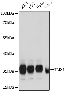
Western blot analysis of various lysates using TMX1 Rabbit pAb (CAB14936) at 1:1000 dilution. Secondary antibody: HRP-conjugated Goat anti-Rabbit IgG (H+L) (CABS014) at 1:10000 dilution. Lysates/proteins: 25μg per lane. Blocking buffer: 3% nonfat dry milk in TBST. Detection: ECL Basic Kit (AbGn00020). Exposure time: 60s.
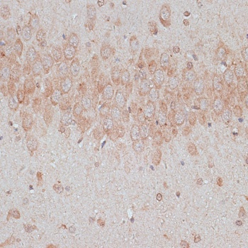
Immunohistochemistry analysis of paraffin-embedded Rat brain using TMX1 Rabbit pAb (CAB14936) at dilution of 1:100 (40x lens). Microwave antigen retrieval performed with 0.01M PBS Buffer (pH 7.2) prior to IHC staining.
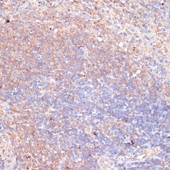
Immunohistochemistry analysis of paraffin-embedded Mouse spleen using TMX1 Rabbit pAb (CAB14936) at dilution of 1:100 (40x lens). Microwave antigen retrieval performed with 0.01M PBS Buffer (pH 7.2) prior to IHC staining.
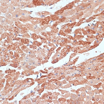
Immunohistochemistry analysis of paraffin-embedded Mouse heart using TMX1 Rabbit pAb (CAB14936) at dilution of 1:100 (40x lens). Microwave antigen retrieval performed with 0.01M PBS Buffer (pH 7.2) prior to IHC staining.