The TNC Antibody (CAB18156) is a high-quality antibody developed for reliable detection and analysis of target proteins. This rabbit polyclonal antibody is highly specific and sensitive, making it ideal for use in experiments such as Western blotting and immunohistochemistry.TNC plays a critical role in cell adhesion, migration, and proliferation, making it a key player in processes like development, tissue repair, and cancer progression. By targeting TNC with this antibody, researchers can effectively study its expression, localization, and function in different cell types and tissues.
This antibody is validated for use in WB, IHC-P, IF/ICC, ELISA applications and has demonstrated reactivity against Human, Mouse, Rat samples.
Product Name:
TNC Antibody
SKU:
CAB18156
Size:
20μL, 100μL
Reactivity:
Human, Mouse, Rat
Immunogen:
Recombinant protein (or fragment).This information is considered to be commercially sensitive.
This gene encodes an extracellular matrix protein with a spatially and temporally restricted tissue distribution. This protein is homohexameric with disulfide-linked subunits, and contains multiple EGF-like and fibronectin type-III domains. It is implicated in guidance of migrating neurons as well as axons during development, synaptic plasticity, and neuronal regeneration.
Purification Method
Affinity purification
Gene ID
3371
RRID
AB_2861946
Buffer Information
Store at -20℃. Avoid freeze / thaw cycles. Buffer: PBS containing 50% glycerol, preserved with proclin300 or sodium azide, pH 7.3.
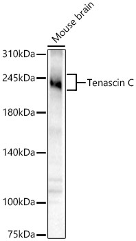
Western blot analysis of lysates from Mouse brain using Tenascin C Rabbit pAb (CAB18156) at 1:400 dilution. Secondary antibody: HRP-conjugated Goat anti-Rabbit IgG (H+L) (CABS014) at 1:10000 dilution. Lysates/proteins: 25 μg per lane. Blocking buffer: 3% nonfat dry milk in TBST. Detection: ECL Basic Kit (AbGn00020). Exposure time: 180s.
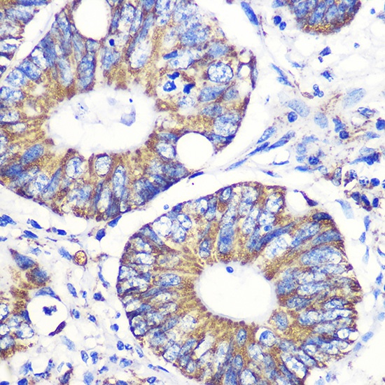
Immunohistochemistry analysis of paraffin-embedded Human colon carcinoma using Tenascin C Rabbit pAb (CAB18156) at dilution of 1:100 (40x lens). Microwave antigen retrieval performed with 0.01M PBS Buffer (pH 7.2) prior to IHC staining.
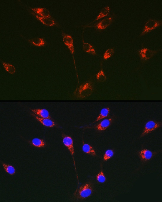
Immunofluorescence analysis of U-87MG cells using Tenascin C Rabbit pAb (CAB18156) at dilution of 1:50 (40x lens). Secondary antibody: Cy3-conjugated Goat anti-Rabbit IgG (H+L) (CABS007) at 1:500 dilution. Blue: DAPI for nuclear staining.