The TNFAIP3 Antibody (CAB2127) is a high-quality antibody developed for reliable detection and analysis of target proteins. This polyclonal antibody, produced in rabbits, exhibits high reactivity with human samples and has been validated for use in applications such as Western blotting.TNFAIP3, also known as A20, is a crucial anti-inflammatory protein that plays a vital role in dampening immune responses and maintaining immune homeostasis. Dysregulation of TNFAIP3 has been implicated in various inflammatory diseases, making it an important target for research in immunology and inflammation-related disorders.
This antibody is validated for use in WB, IHC-P, ELISA applications and has demonstrated reactivity against Human, Rat samples.
Product Name:
TNFAIP3 Antibody
SKU:
CAB2127
Size:
20μL, 100μL
Reactivity:
Human, Rat
Conjugate:
Unconjugated
Immunogen:
Recombinant protein (or fragment).This information is considered to be commercially sensitive.
Recommended starting concentration is 1 μg/mL. Please optimize the concentration based on your specific assay requirements.
Synonyms:
A20, AISBL, AIFBL1, OTUD7C, TNFA1P2, TNFAIP3
Positive Sample:
THP-1 treated with TPA and LPS, Rat thymus
Cellular Localization:
Cytoplasm, Lysosome, Nucleus.
Calculated MW:
90kDa
Observed MW:
90kDa
This gene was identified as a gene whose expression is rapidly induced by the tumor necrosis factor (TNF). The protein encoded by this gene is a zinc finger protein and ubiqitin-editing enzyme, and has been shown to inhibit NF-kappa B activation as well as TNF-mediated apoptosis. The encoded protein, which has both ubiquitin ligase and deubiquitinase activities, is involved in the cytokine-mediated immune and inflammatory responses. Several transcript variants encoding the same protein have been found for this gene.
Purification Method
Affinity purification
Gene ID
7128
RRID
AB_2764146
Buffer Information
Store at -20℃. Avoid freeze / thaw cycles. Buffer: PBS containing 50% glycerol, preserved with proclin300 or sodium azide, pH 7.3.
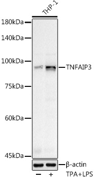
Western blot analysis of various lysates using (CAB2127) at 1:500 dilution. THP-1 cells were treated with PMA/TPA (200 nM) at 37℃ for 15 minutes after serum-starvation overnight. THP-1 cells were treated with LPS (1 μg/ml) at 37℃ for 8 hours. Secondary antibody: HRP-conjugated Goat anti-Rabbit IgG (H+L) (CABS014) at 1:10000 dilution. Lysates/proteins: 25μg per lane. Blocking buffer: 3% nonfat dry milk in TBST. Detection: ECL Basic Kit (AbGn00020). Exposure time: 40s.
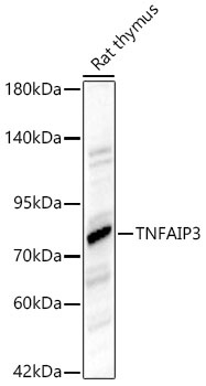
Western blot analysis of lysates from Rat thymus using TNFAIP3 Rabbit pAb (CAB2127) at 1:1000 dilution. Secondary antibody: HRP-conjugated Goat anti-Rabbit IgG (H+L) (CABS014) at 1:10000 dilution. Lysates/proteins: 25 μg per lane. Blocking buffer: 3% nonfat dry milk in TBST. Detection: ECL Basic Kit (AbGn00020). Exposure time:20s.
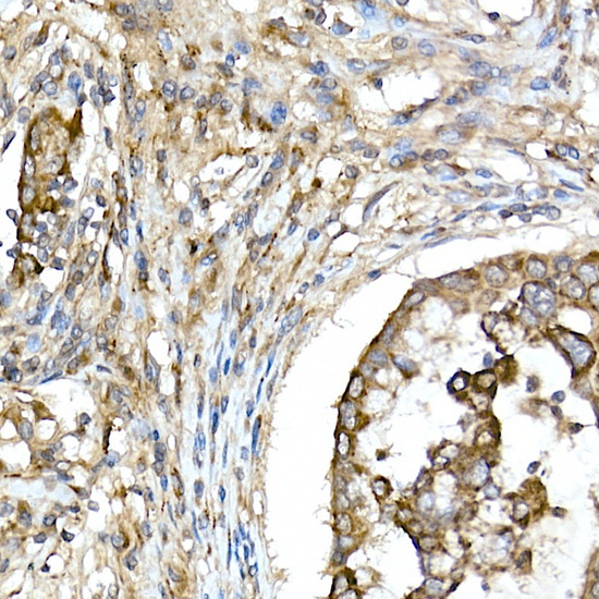
Immunohistochemistry analysis of paraffin-embedded Human lung cancer using TNFAIP3 Rabbit pAb (CAB2127) at dilution of 1:50 (40x lens). High pressure antigen retrieval performed with 0.01M Citrate buffer (pH 6.0) prior to IHC staining.
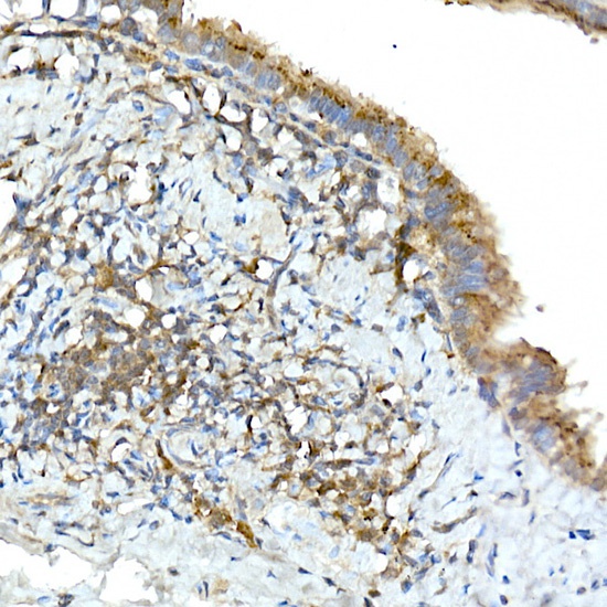
Immunohistochemistry analysis of paraffin-embedded Rat lung using TNFAIP3 Rabbit pAb (CAB2127) at dilution of 1:50 (40x lens). High pressure antigen retrieval performed with 0.01M Citrate buffer (pH 6.0) prior to IHC staining.


")


