The TNFRSF10D Antibody (CAB6136) is a high-quality antibody developed for reliable detection and analysis of target proteins. This antibody, generated in rabbits, has high reactivity with human samples and is validated for use in various applications, including Western blotting. By binding specifically to the TNFRSF10D protein, this antibody enables accurate detection and analysis in a variety of cell types, making it well-suited for immunology and cancer research studies.
This antibody is validated for use in WB, IHC-P, ELISA applications and has demonstrated reactivity against Human, Mouse samples.
Product Name:
TNFRSF10D Antibody
SKU:
CAB6136
Size:
20μL, 100μL
Reactivity:
Human, Mouse
Conjugate:
Unconjugated
Immunogen:
Recombinant protein (or fragment).This information is considered to be commercially sensitive.
Recommended starting concentration is 1 μg/mL. Please optimize the concentration based on your specific assay requirements.
Synonyms:
DCR2, CD264, TRUNDD, TRAILR4, TRAIL-R4, TNFRSF10D
Positive Sample:
THP-1
Cellular Localization:
Membrane, Single-Pass Type I Membrane Protein.
Calculated MW:
42kDa
Observed MW:
47kDa
The protein encoded by this gene is a member of the TNF-receptor superfamily. This receptor contains an extracellular TRAIL-binding domain, a transmembrane domain, and a truncated cytoplamic death domain. This receptor does not induce apoptosis, and has been shown to play an inhibitory role in TRAIL-induced cell apoptosis.
Purification Method
Affinity purification
Gene ID
8793
RRID
AB_2766766
Buffer Information
Store at -20℃. Avoid freeze / thaw cycles. Buffer: PBS containing 50% glycerol, preserved with proclin300 or sodium azide, pH 7.3.
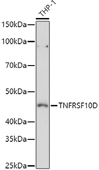
Western blot analysis of lysates from THP-1 cells, using TNFRSF10D Rabbit pAb (CAB6136) at 1:1000 dilution. Secondary antibody: HRP-conjugated Goat anti-Rabbit IgG (H+L) (CABS014) at 1:10000 dilution. Lysates/proteins: 25μg per lane. Blocking buffer: 3% nonfat dry milk in TBST. Detection: ECL Basic Kit (AbGn00020). Exposure time: 60s.
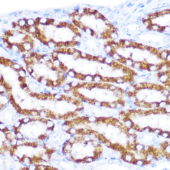
Immunohistochemistry analysis of paraffin-embedded Mouse kidney using TNFRSF10D Rabbit pAb (CAB6136) at dilution of 1:100 (40x lens). Microwave antigen retrieval performed with 0.01M Tris/EDTA Buffer (pH 9.0) prior to IHC staining.