The TNFR1/TNFRSF1A Antibody (CAB1540) is a high-quality antibody developed for reliable detection and analysis of target proteins. This antibody, produced in rabbits, exhibits high reactivity with human samples and is validated for use in Western blot applications. By binding specifically to the TNFRSF1A protein, this antibody enables accurate detection and analysis in a variety of cell types, making it an ideal choice for studies in immunology and inflammation-related research.TNFRSF1A, also known as TNF receptor superfamily member 1A, is a key player in the pro-inflammatory signaling pathways, making it a target of interest for research related to inflammatory diseases, autoimmune disorders, and cancer.
This antibody is validated for use in WB, IHC-P, IF/ICC, ELISA applications and has demonstrated reactivity against Human, Mouse, Rat samples.
Product Name:
TNFR1/TNFRSF1A Antibody
SKU:
CAB1540
Size:
20μL, 100μL
Reactivity:
Human, Mouse, Rat
Conjugate:
Unconjugated
Immunogen:
Recombinant protein (or fragment).This information is considered to be commercially sensitive.
Cell Membrane, Golgi Apparatus Membrane, Secreted, Secreted, Single-Pass Type I Membrane Protein.
Calculated MW:
50kDa
Observed MW:
55kDa
This gene encodes a member of the TNF receptor superfamily of proteins. The encoded receptor is found in membrane-bound and soluble forms that interact with membrane-bound and soluble forms, respectively, of its ligand, tumor necrosis factor alpha. Binding of membrane-bound tumor necrosis factor alpha to the membrane-bound receptor induces receptor trimerization and activation, which plays a role in cell survival, apoptosis, and inflammation. Proteolytic processing of the encoded receptor results in release of the soluble form of the receptor, which can interact with free tumor necrosis factor alpha to inhibit inflammation. Mutations in this gene underlie tumor necrosis factor receptor-associated periodic syndrome (TRAPS), characterized by fever, abdominal pain and other features. Mutations in this gene may also be associated with multiple sclerosis in human patients.
Purification Method
Affinity purification
Gene ID
7132
RRID
AB_2762307
Buffer Information
Store at -20℃. Avoid freeze / thaw cycles. Buffer: PBS containing 50% glycerol, preserved with proclin300 or sodium azide, pH 7.3.
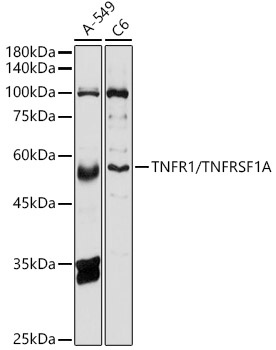
Western blot analysis of various lysates using TNFR1/TNFRSF1A Rabbit pAb (CAB1540) at 1:500 dilution. Secondary antibody: HRP-conjugated Goat anti-Rabbit IgG (H+L) (CABS014) at 1:10000 dilution. Lysates/proteins: 25μg per lane. Blocking buffer: 3% nonfat dry milk in TBST. Detection: ECL Basic Kit (AbGn00020). Exposure time: 10s.
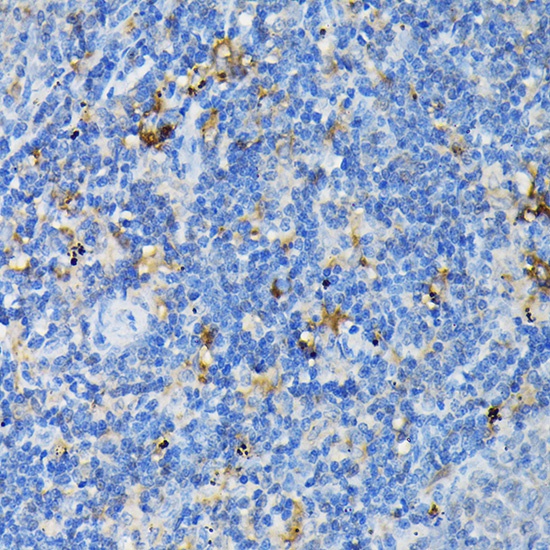
Immunohistochemistry analysis of paraffin-embedded Rat spleen using TNFR1/TNFRSF1A Rabbit pAb (CAB1540) at dilution of 1:100 (40x lens). High pressure antigen retrieval performed with 0.01M Citrate buffer (pH 6.0) prior to IHC staining.


")
")
")
")
