The TNFRSF6B Antibody (CAB0649) is a high-quality antibody developed for reliable detection and analysis of target proteins. This antibody, raised in rabbits, has high reactivity with human samples and is validated for use in Western blot applications. By binding to TNFRSF6B, this antibody enables accurate detection and analysis of the protein in various cell types, making it ideal for studies in immunology, cancer research, and other related fields.TNFRSF6B is involved in the regulation of immune responses and apoptosis, playing a crucial role in modulating inflammatory processes and cell survival. Its role as a decoy receptor for certain cytokines and apoptosis-inducing ligands gives it significant importance in diseases such as cancer, autoimmune disorders, and inflammatory conditions.
This antibody is validated for use in WB, ELISA applications and has demonstrated reactivity against Human, Rat samples.
Product Name:
TNFRSF6B Antibody
SKU:
CAB0649
Size:
20μL, 100μL
Reactivity:
Human, Rat
Conjugate:
Unconjugated
Immunogen:
Recombinant protein (or fragment).This information is considered to be commercially sensitive.
Recommended starting concentration is 1 μg/mL. Please optimize the concentration based on your specific assay requirements.
Synonyms:
M68, TR6, DCR3, M68E, DJ583P15.1.1, TNFRSF6B
Positive Sample:
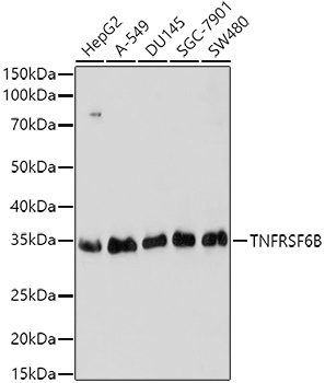
HepG2, A-549, DU145, SGC-7901, SW480
Cellular Localization:
Secreted.
Calculated MW:
33kDa
Observed MW:
33kDa
This gene belongs to the tumor necrosis factor receptor superfamily. The encoded protein is postulated to play a regulatory role in suppressing FasL- and LIGHT-mediated cell death. It acts as a decoy receptor that competes with death receptors for ligand binding. Over-expression of this gene has been noted in gastrointestinal tract tumors. Read-through transcription into this gene from the neighboring upstream gene, which encodes regulator of telomere elongation helicase 1 (RTEL1), generates a non-coding transcript.
Purification Method
Affinity purification
Gene ID
8771
RRID
AB_2757315
Buffer Information
Store at -20℃. Avoid freeze / thaw cycles. Buffer: PBS with 0.09% Sodium azide,50% glycerol,pH7.3.
Western blot analysis of various lysates using TNFRSF6B Rabbit pAb (CAB0649) at 1:1000 dilution. Secondary antibody: HRP-conjugated Goat anti-Rabbit IgG (H+L) (CABS014) at 1:10000 dilution. Lysates/proteins: 25μg per lane. Blocking buffer: 3% nonfat dry milk in TBST. Detection: ECL Basic Kit (AbGn00020). Exposure time: 60s.