The TNPO1 Antibody (CAB9435) is a high-quality antibody developed for reliable detection and analysis of target proteins. This antibody, produced in rabbits, exhibits high reactivity with human samples and has been validated for use in Western blotting applications. By targeting the Transportin-1 protein, this antibody enables researchers to study its role in the transport of macromolecules across the nuclear membrane in different cell types, making it an essential tool for studies in molecular biology and cell biology.Transportin-1 is a key player in the regulation of cellular processes such as gene expression, signal transduction, and protein transport.
This antibody is validated for use in WB, ELISA applications and has demonstrated reactivity against Human, Mouse, Rat samples.
Product Name:
TNPO1 Antibody
SKU:
CAB9435
Size:
20μL, 100μL
Reactivity:
Human, Mouse, Rat
Conjugate:
Unconjugated
Immunogen:
Synthetic peptide. This information is considered to be commercially sensitive.
Recommended starting concentration is 1 μg/mL. Please optimize the concentration based on your specific assay requirements.
Synonyms:
MIP, TRN, IPO2, MIP1, KPNB2, TNPO1
Positive Sample:
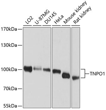
LO2, U-87MG, DU145, HeLa, Mouse kidney, Rat kidney
Cellular Localization:
Cytoplasm, Nucleus.
Calculated MW:
102kDa
Observed MW:
85-102kDa
This gene encodes the beta subunit of the karyopherin receptor complex which interacts with nuclear localization signals to target nuclear proteins to the nucleus. The karyopherin receptor complex is a heterodimer of an alpha subunit which recognizes the nuclear localization signal and a beta subunit which docks the complex at nucleoporins. Alternate splicing of this gene results in several transcript variants encoding different proteins.
Purification Method
Affinity purification
Gene ID
3842
RRID
AB_2772672
Buffer Information
Store at -20℃. Avoid freeze / thaw cycles. Buffer: PBS containing 50% glycerol, preserved with proclin300 or sodium azide, pH 7.3.
Western blot analysis of various lysates using TNPO1 Rabbit pAb (CAB9435) at 1:1000 dilution. Secondary antibody: HRP-conjugated Goat anti-Rabbit IgG (H+L) (CABS014) at 1:10000 dilution. Lysates/proteins: 25μg per lane. Blocking buffer: 3% nonfat dry milk in TBST. Detection: ECL Enhanced Kit (AbGn00021). Exposure time: 1s.