The TNXB Antibody (CAB2535) is a high-quality antibody developed for reliable detection and analysis of target proteins. This antibody, produced in rabbits, specifically targets tenascin-X and is highly reactive with human samples. Validated for use in Western blot applications, it allows for the detection and analysis of tenascin-X in a variety of cell types.Tenascin-X is a crucial component of the extracellular matrix, playing a key role in tissue development and wound healing. Aberrant expression of tenascin-X has been implicated in various diseases, including Ehlers-Danlos syndrome and certain types of cancer.
This antibody is validated for use in WB, ELISA applications and has demonstrated reactivity against Mouse, Rat samples.
Product Name:
TNXB Antibody
SKU:
CAB2535
Size:
20μL, 100μL
Reactivity:
Mouse, Rat
Conjugate:
Unconjugated
Immunogen:
Recombinant protein (or fragment).This information is considered to be commercially sensitive.
This gene encodes a member of the tenascin family of extracellular matrix glycoproteins. The tenascins have anti-adhesive effects, as opposed to fibronectin which is adhesive. This protein is thought to function in matrix maturation during wound healing, and its deficiency has been associated with the connective tissue disorder Ehlers-Danlos syndrome. This gene localizes to the major histocompatibility complex (MHC) class III region on chromosome 6. It is one of four genes in this cluster which have been duplicated. The duplicated copy of this gene is incomplete and is a pseudogene which is transcribed but does not encode a protein. The structure of this gene is unusual in that it overlaps the CREBL1 and CYP21A2 genes at its 5' and 3' ends, respectively. Multiple transcript variants encoding different isoforms have been found for this gene.
Purification Method
Affinity purification
Gene ID
7148
RRID
AB_2764425
Buffer Information
Store at -20℃. Avoid freeze / thaw cycles. Buffer: PBS with 0.01% thimerosal,50% glycerol,pH7.3.
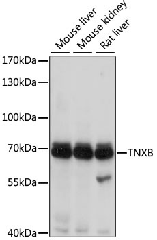
Western blot analysis of various lysates using TNXB Rabbit pAb (CAB2535) at 1:1000 dilution. Secondary antibody: HRP-conjugated Goat anti-Rabbit IgG (H+L) (CABS014) at 1:10000 dilution. Lysates/proteins: 25μg per lane. Blocking buffer: 3% nonfat dry milk in TBST. Detection: ECL Basic Kit (AbGn00020). Exposure time: 1s.