The TOM20 Monoclonal Antibody (CAB19403) is a high-quality antibody developed for reliable detection and analysis of target proteins. Enables protein-transporting ATPase activity and unfolded protein binding activity. Involved in protein targeting to mitochondrion. Located in mitochondria-associated endoplasmic reticulum membrane and mitochondrial outer membrane. RRID AB_2862646 Gene ID 9804 Swiss Prot Synonym MAS20; MOM19; TOM20
This antibody is validated for use in WB, IHC-P, IF/ICC, IP, ELISA applications and has demonstrated reactivity against Human, Mouse, Rat samples.
Product Name:
TOM20 Monoclonal Antibody
SKU:
CAB19403
Size:
100μL, 20μL
Reactivity:
Human, Mouse, Rat
Clone Number:
ARC5002-01
Conjugate:
Unconjugated
Immunogen:
Recombinant protein (or fragment).This information is considered to be commercially sensitive.
Tested Applications:
WBIHC-PIF/ICCIPELISA
Recommended Dilution:
WB
1:5000 - 1:160000
IHC-P
1:1000 - 1:5000
IF
/
ICC
1:100 - 1:2000
IP
0.5μg-4μg antibody for 200μg-400μg extracts of whole cells
ELISA
Recommended starting concentration is 1 μg/mL. Please optimize the concentration based on your specific assay requirements.
Enables protein-transporting ATPase activity and unfolded protein binding activity. Involved in protein targeting to mitochondrion. Located in mitochondria-associated endoplasmic reticulum membrane and mitochondrial outer membrane. RRID AB_2862646 Gene ID 9804 Swiss Prot Synonym MAS20; MOM19; TOM20
Purification Method:
Affinity purification
Gene ID:
9804
RRID:
AB_2862646
Buffer Information:
Store at -20℃. Avoid freeze / thaw cycles. Buffer: PBS with 0.09% sodium azide,0.05% BSA,50% glycerol,pH7.3.
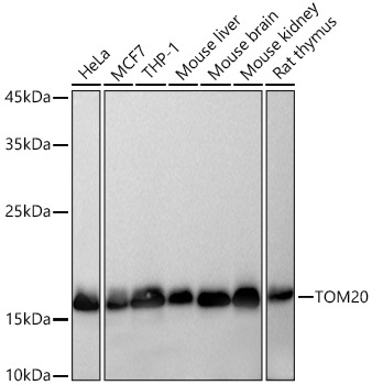
Western blot analysis of various lysates using TOM20 Rabbit mAb (CAB19403) at 1:5000 dilution incubated overnight at 4℃. Secondary antibody: HRP-conjugated Goat anti-Rabbit IgG (H+L) (AS014) at 1:10000 dilution. Lysates/proteins: 25 μg per lane. Blocking buffer: 3% nonfat dry milk in TBST. Detection: ECL Basic Kit (AbGn00020). Exposure time: 10s.
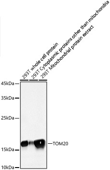
Western blot analysis of lysates from 293T cells using TOM20 Rabbit mAb (CAB19403) at 1:5000 dilution incubated overnight at 4℃. Secondary antibody: HRP-conjugated Goat anti-Rabbit IgG (H+L) (AS014) at 1:10000 dilution. Lysates/proteins: 25 μg per lane. Blocking buffer: 3% nonfat dry milk in TBST. Detection: ECL Basic Kit (AbGn00020). Exposure time: 10s.
Immunohistochemistry analysis of paraffin-embedded Mouse lung tissue using TOM20 Rabbit mAb (CAB19403) at a dilution of 1:5000 (40x lens). High pressure antigen retrieval performed with 0.01M Tris-EDTA Buffer (pH 9.0) prior to IHC staining.
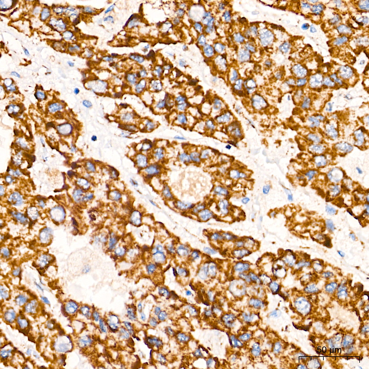
Immunohistochemistry analysis of paraffin-embedded Human liver cancer tissue using TOM20 Rabbit mAb (CAB19403) at a dilution of 1:5000 (40x lens). High pressure antigen retrieval performed with 0.01M Tris-EDTA Buffer (pH 9.0) prior to IHC staining.
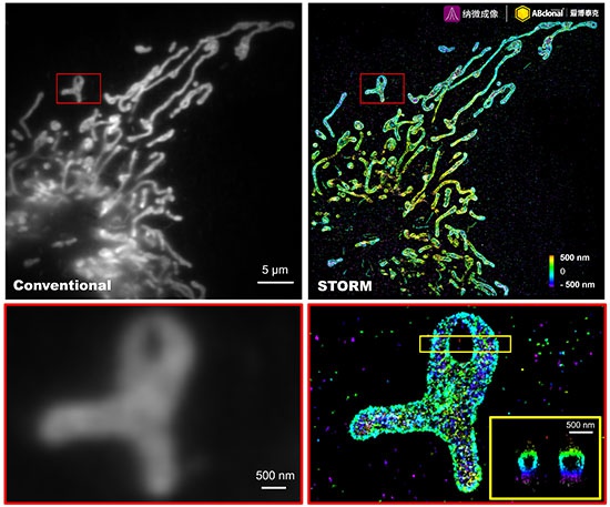
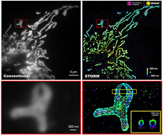
The STORM super-resolution (SR) imaging of U-2 OS cells using TOM20 Rabbit mAb (CAB19403, ABclonal) at dilution of 1:100 with 3% paraformaldehyde (PFA) +0.1% glutaraldehyde (GA) fixation. The immunostaining was performed by Full Automatic Immunofluorescence Workflow System (Workflow Ultra300, Nano-Micro imaging, China). Image was performed with Single-Molecule Localization Super-Resolution Microscopy (STORM Ultra300, Nano-Micro imaging, China). We acknowledge Ningbo Nano-Micro imaging Biotechnology Co., Ltd. (宁波纳微成像生物科技有限公司) in SR image processing and kindly providing this image.
The STORM super-resolution (SR) imaging of U-2 OS cells using TOM20 Rabbit mAb (CAB19403, ABclonal) at dilution of 1:200 with 3% paraformaldehyde (PFA) +0.1% glutaraldehyde (GA) fixation. The immunostaining was performed by Full Automatic Immunofluorescence Workflow System (Workflow Ultra300, Nano-Micro imaging, China). Image was performed with Single-Molecule Localization Super-Resolution Microscopy (STORM Ultra300, Nano-Micro imaging, China). We acknowledge Nano-Micro imaging Biotechnology Co., Ltd. in SR image processing and kindly providing this image.
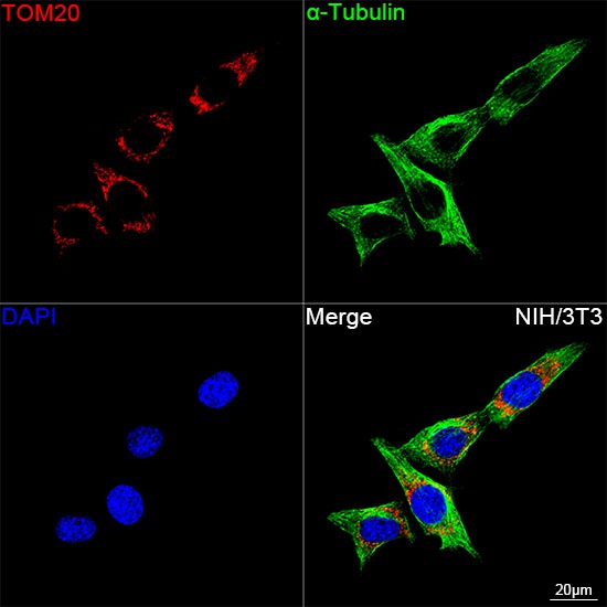
Confocal imaging of NIH/3T3 cells using TOM20 Rabbit mAb (CAB19403, dilution 1:2000) followed by a further incubation with Cy3 Goat Anti-Rabbit IgG (H+L) (AS007, dilution 1:500) (Red). The cells were counterstained with α-Tubulin Mouse mAb (AC012, dilution 1:400) followed by incubation with ABflo® 488-conjugated Goat Anti-Mouse IgG (H+L) Ab (AS076, dilution 1:500) (Green). DAPI was used for nuclear staining (Blue). Objective: 100x.
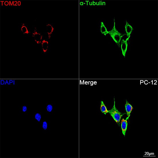
Confocal imaging of PC-12 cells using TOM20 Rabbit mAb (CAB19403, dilution 1:2000) followed by a further incubation with Cy3 Goat Anti-Rabbit IgG (H+L) (AS007, dilution 1:500) (Red). The cells were counterstained with α-Tubulin Mouse mAb (AC012, dilution 1:400) followed by incubation with ABflo® 488-conjugated Goat Anti-Mouse IgG (H+L) Ab (AS076, dilution 1:500) (Green). DAPI was used for nuclear staining (Blue). Objective: 100x.
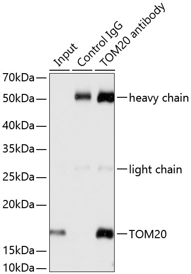
Immunoprecipitation analysis of 200 μg extracts from HeLa cells using 3 μg TOM20 antibody (CAB19403). Western blot was performed from the immunoprecipitate using TOM20 antibody (CAB19403) at a dilution of 1:1000.