The TOM20 Antibody (CAB6774) is a high-quality antibody developed for reliable detection and analysis of target proteins. Enables protein-transporting ATPase activity and unfolded protein binding activity. Involved in protein targeting to mitochondrion. Located in mitochondria-associated endoplasmic reticulum membrane and mitochondrial outer membrane. RRID AB_2767357 Gene ID 9804 Swiss Prot Synonym MAS20; MOM19; TOM20
This antibody is validated for use in WB, IHC-P, IF/ICC, IP, ELISA applications and has demonstrated reactivity against Human, Mouse, Rat samples.
Product Name:
TOM20 Antibody
SKU:
CAB6774
Size:
100μL, 20μL
Reactivity:
Human, Mouse, Rat
Clone Number:
-
Conjugate:
Unconjugated
Immunogen:
Recombinant protein (or fragment).This information is considered to be commercially sensitive.
Tested Applications:
WBIHC-PIF/ICCIPELISA
Recommended Dilution:
WB
1:500 - 1:2000
IHC-P
1:50 - 1:200
IF
/
ICC
1:50 - 1:200
IP
0.5μg-4μg antibody for 200μg-400μg extracts of whole cells
ELISA
Recommended starting concentration is 1 μg/mL. Please optimize the concentration based on your specific assay requirements.
Synonyms:
MAS20, MOM19, TOM20
Positive Sample:
HeLa, SH-SY5Y, 293T, Mouse brain, Mouse kidney, Rat brain
Enables protein-transporting ATPase activity and unfolded protein binding activity. Involved in protein targeting to mitochondrion. Located in mitochondria-associated endoplasmic reticulum membrane and mitochondrial outer membrane. RRID AB_2767357 Gene ID 9804 Swiss Prot Synonym MAS20; MOM19; TOM20
Purification Method:
Affinity purification
Gene ID:
9804
RRID:
AB_2767357
Buffer Information:
Store at -20℃. Avoid freeze / thaw cycles. Buffer: PBS containing 50% glycerol, preserved with proclin300 or sodium azide, pH 7.3.
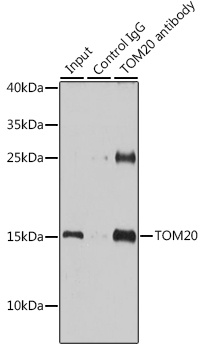
Immunoprecipitation analysis of 200 μg extracts of HeLa cells using 3 μg TOM20 antibody (CAB6774). Western blot was performed from the immunoprecipitate using TOM20 antibody (CAB6774) at a dilution of 1:1000.
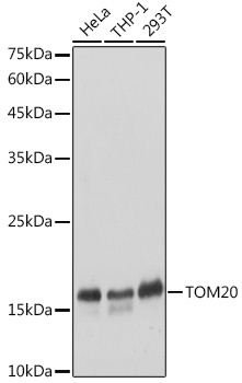
Western blot analysis of various lysates using [KO Validated] TOM20 Rabbit pAb (CAB6774) at 1:3000 dilution. Secondary antibody: HRP-conjugated Goat anti-Rabbit IgG (H+L) (AS014) at 1:10000 dilution. Lysates/proteins: 25μg per lane. Blocking buffer: 3% nonfat dry milk in TBST. Detection: ECL Basic Kit (AbGn00020). Exposure time: 1s.
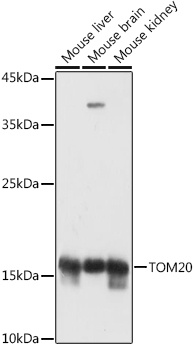
Western blot analysis of various lysates using [KD Validated] TOM20 Rabbit pAb (CAB6774) at 1:1000 dilution. Secondary antibody: HRP-conjugated Goat anti-Rabbit IgG (H+L) (AS014) at 1:10000 dilution. Lysates/proteins: 25μg per lane. Blocking buffer: 3% nonfat dry milk in TBST. Detection: ECL Basic Kit (AbGn00020). Exposure time: 1s.
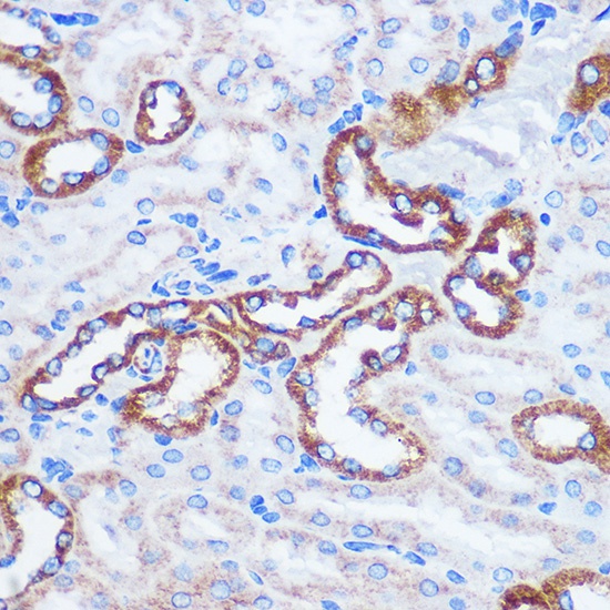
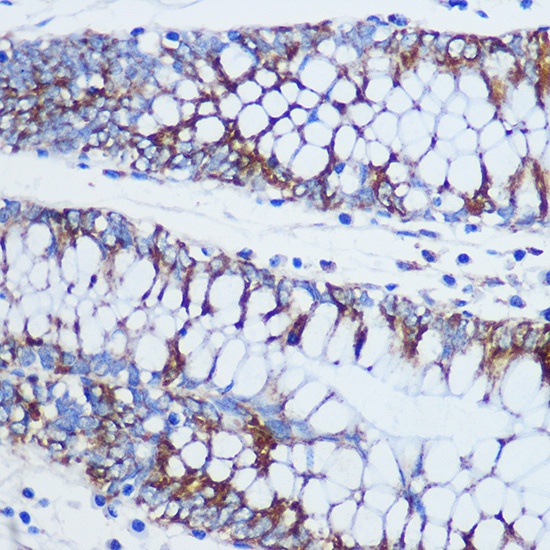
Immunohistochemistry analysis of paraffin-embedded Mouse kidney using TOM20 Rabbit pAb (CAB6774) at dilution of 1:100 (40x lens). Microwave antigen retrieval performed with 0.01M PBS Buffer (pH 7.2) prior to IHC staining.
Immunohistochemistry analysis of paraffin-embedded Human colon using TOM20 Rabbit pAb (CAB6774) at dilution of 1:100 (40x lens). Microwave antigen retrieval performed with 0.01M PBS Buffer (pH 7.2) prior to IHC staining.
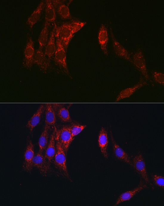
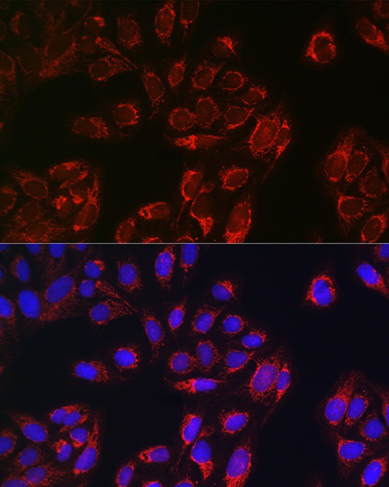
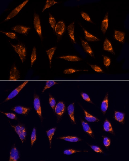
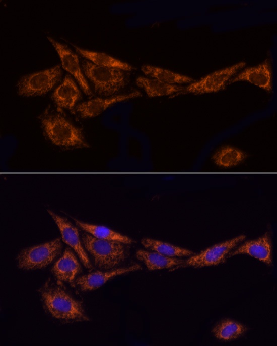
Immunofluorescence analysis of C6 cells using [KO Validated] TOM20 Rabbit pAb (CAB6774) at dilution of 1:100 (40x lens). Secondary antibody: Cy3-conjugated Goat anti-Rabbit IgG (H+L) (AS007) at 1:500 dilution. Blue: DAPI for nuclear staining.
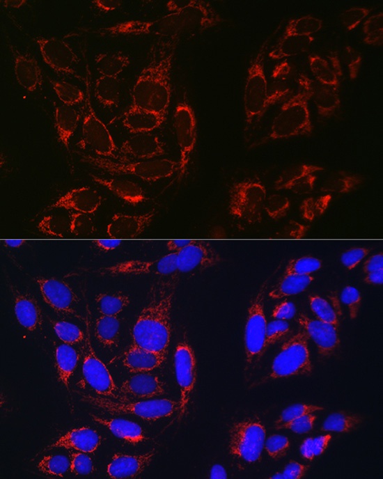
Immunofluorescence analysis of U-2 OS cells using [KO Validated] TOM20 Rabbit pAb (CAB6774) at dilution of 1:100 (40x lens). Secondary antibody: Cy3-conjugated Goat anti-Rabbit IgG (H+L) (AS007) at 1:500 dilution. Blue: DAPI for nuclear staining.
Immunofluorescence analysis of C6 cells using [KO Validated] TOM20 Rabbit pAb (CAB6774) at dilution of 1:100 (40x lens). Secondary antibody: Cy3-conjugated Goat anti-Rabbit IgG (H+L) (AS007) at 1:500 dilution. Blue: DAPI for nuclear staining.
Immunofluorescence analysis of U2OS cells using [KO Validated] TOM20 Rabbit pAb (CAB6774) at dilution of 1:100 (40x lens). Secondary antibody: Cy3-conjugated Goat anti-Rabbit IgG (H+L) (AS007) at 1:500 dilution. Blue: DAPI for nuclear staining.
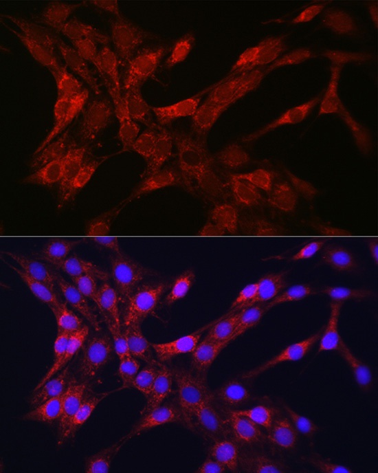
Immunofluorescence analysis of HeLa cells using TOM20 Rabbit pAb (CAB6774) at dilution of 1:200 (40x lens). Secondary antibody: Cy3-conjugated Goat anti-Rabbit IgG (H+L) (AS007) at 1:500 dilution. Blue: DAPI for nuclear staining.
Immunofluorescence analysis of PC-12 cells using TOM20 Rabbit pAb (CAB6774) at dilution of 1:200 (40x lens). Secondary antibody: Cy3-conjugated Goat anti-Rabbit IgG (H+L) (AS007) at 1:500 dilution. Blue: DAPI for nuclear staining.


")
