The TPX2 Antibody (CAB18327) is a high-quality antibody developed for reliable detection and analysis of target proteins. This rabbit polyclonal antibody is specifically optimized for use in various applications, including Western blot, immunofluorescence, immunoprecipitation, and flow cytometry.TPX2 is a well-known regulator of microtubule organization and plays a crucial role in ensuring proper chromosome segregation during cell division. Dysregulation of TPX2 expression has been linked to cancer development and progression, making it a promising target for cancer research and drug development.
This antibody is validated for use in WB, IHC-P, IF/ICC, ELISA applications and has demonstrated reactivity against Human, Rat samples.
Product Name:
TPX2 Antibody
SKU:
CAB18327
Size:
20μL, 100μL
Reactivity:
Human, Rat
Immunogen:
Recombinant protein (or fragment).This information is considered to be commercially sensitive.
Cytoplasm, Nucleus, Cytoskeleton, Spindle, Spindle Pole.
Calculated MW:
86kDa
Observed MW:
105kDa
Enables importin-alpha family protein binding activity and protein kinase binding activity. Involved in activation of protein kinase activity; microtubule cytoskeleton organization; and negative regulation of microtubule depolymerization. Located in intercellular bridge; mitotic spindle; and nucleoplasm. Colocalizes with spindle pole.
Purification Method
Affinity purification
Gene ID
22974
RRID
AB_2862099
Buffer Information
Store at -20℃. Avoid freeze / thaw cycles. Buffer: PBS containing 50% glycerol, preserved with proclin300 or sodium azide, pH 7.3.
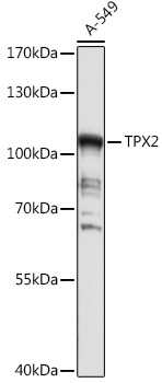
Western blot analysis of lysates from A-549 cells, using TPX2 Rabbit pAb (CAB18327) at 1:1000 dilution. Secondary antibody: HRP-conjugated Goat anti-Rabbit IgG (H+L) (CABS014) at 1:10000 dilution. Lysates/proteins: 25μg per lane. Blocking buffer: 3% nonfat dry milk in TBST. Detection: ECL Basic Kit (AbGn00020). Exposure time: 30s.
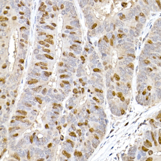
Immunohistochemistry analysis of paraffin-embedded Human colon carcinoma using TPX2 Rabbit pAb (CAB18327) at dilution of 1:100 (40x lens). High pressure antigen retrieval performed with 0.01M Citrate buffer (pH 6.0) prior to IHC staining.
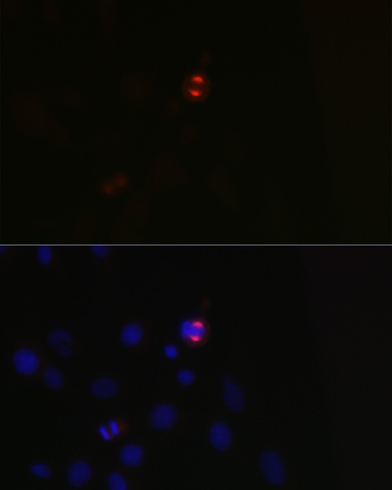
Immunofluorescence analysis of C6 cells using TPX2 Rabbit pAb (CAB18327) at dilution of 1:100 (40x lens). Secondary antibody: Cy3-conjugated Goat anti-Rabbit IgG (H+L) (CABS007) at 1:500 dilution. Blue: DAPI for nuclear staining.
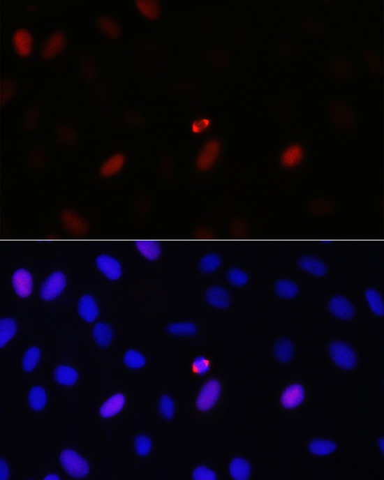
Immunofluorescence analysis of U-2 OS cells using TPX2 Rabbit pAb (CAB18327) at dilution of 1:100 (40x lens). Secondary antibody: Cy3-conjugated Goat anti-Rabbit IgG (H+L) (CABS007) at 1:500 dilution. Blue: DAPI for nuclear staining.