The TREX1 Antibody (CAB6778) is a high-quality antibody developed for reliable detection and analysis of target proteins. This antibody, produced in rabbits, is highly specific to human samples and has been validated for use in Western blot applications. By binding to the TREX1 protein, this antibody allows for precise detection and analysis in various cell types, making it ideal for studies in immunology, autoimmunity, and cancer research.TREX1, also known as DNase III, plays a crucial role in maintaining genomic stability by clearing out DNA fragments that can trigger inflammatory responses.
This antibody is validated for use in WB, IF/ICC, ELISA applications and has demonstrated reactivity against Human, Mouse, Rat samples.
Product Name:
TREX1 Antibody
SKU:
CAB6778
Size:
20μL, 100μL
Reactivity:
Human, Mouse, Rat
Conjugate:
Unconjugated
Immunogen:
Recombinant protein (or fragment).This information is considered to be commercially sensitive.
This gene encodes a nuclear protein with 3' exonuclease activity. The encoded protein may play a role in DNA repair and serve as a proofreading function for DNA polymerase. Mutations in this gene result in Aicardi-Goutieres syndrome, chilblain lupus, Cree encephalitis, and other diseases of the immune system. Alternative splicing results in multiple transcript variants.
Purification Method
Affinity purification
Gene ID
11277
RRID
AB_2767361
Buffer Information
Store at -20℃. Avoid freeze / thaw cycles. Buffer: PBS containing 50% glycerol, preserved with proclin300 or sodium azide, pH 7.3.
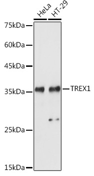
Western blot analysis of various lysates using TREX1 Rabbit pAb (CAB6778) at 1:1000 dilution. Secondary antibody: HRP-conjugated Goat anti-Rabbit IgG (H+L) (CABS014) at 1:10000 dilution. Lysates/proteins: 25μg per lane. Blocking buffer: 3% nonfat dry milk in TBST. Detection: ECL Basic Kit (AbGn00020). Exposure time: 1s.
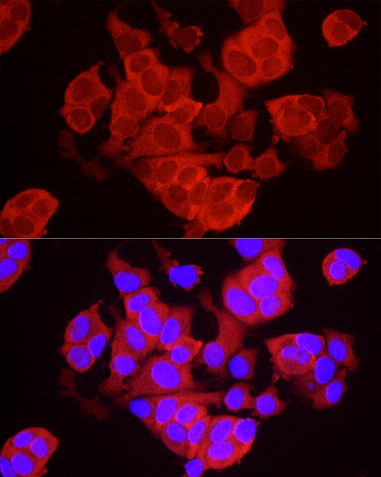
Immunofluorescence analysis of MCF7 cells using TREX1 Rabbit pAb (CAB6778) at dilution of 1:50 (40x lens). Secondary antibody: Cy3-conjugated Goat anti-Rabbit IgG (H+L) (CABS007) at 1:500 dilution. Blue: DAPI for nuclear staining.
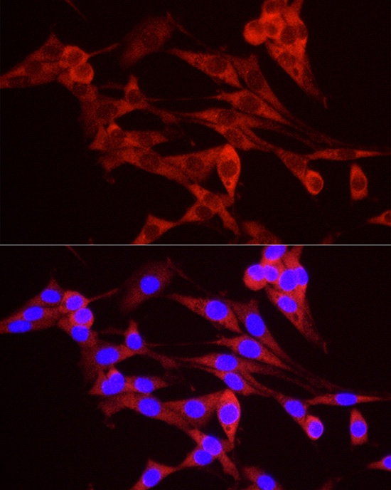
Immunofluorescence analysis of PC-12 cells using TREX1 Rabbit pAb (CAB6778) at dilution of 1:50 (40x lens). Secondary antibody: Cy3-conjugated Goat anti-Rabbit IgG (H+L) (CABS007) at 1:500 dilution. Blue: DAPI for nuclear staining.