The TREX1 Monoclonal Antibody (CAB3819) is a high-quality antibody developed for reliable detection and analysis of target proteins. This antibody is produced in rabbits and is highly specific for detecting TREX1 in human samples, making it an essential tool for studies in immunology and cancer research. TREX1 is known for its role in DNA degradation and its involvement in autoimmune diseases such as Aicardi-Goutières syndrome and systemic lupus erythematosus. By targeting TREX1, researchers can better understand its functions in maintaining genome stability and modulating immune responses, leading to potential insights for therapeutic interventions in various disease conditions.
This antibody is validated for use in WB, IP, ELISA applications and has demonstrated reactivity against Human samples.
Product Name:
TREX1 Monoclonal Antibody
SKU:
CAB3819
Size:
20μL, 100μL
Reactivity:
Human
Clone Number:
ARC0841
Conjugate:
Unconjugated
Immunogen:
Recombinant protein (or fragment).This information is considered to be commercially sensitive.
This gene encodes a nuclear protein with 3' exonuclease activity. The encoded protein may play a role in DNA repair and serve as a proofreading function for DNA polymerase. Mutations in this gene result in Aicardi-Goutieres syndrome, chilblain lupus, Cree encephalitis, and other diseases of the immune system. Alternative splicing results in multiple transcript variants.
Purification Method
Affinity purification
Gene ID
11277
RRID
AB_2863146
Buffer Information
Store at -20℃. Avoid freeze / thaw cycles. Buffer: PBS containing 50% glycerol and 0.05% BSA, preserved with proclin300 or sodium azide, pH 7.3.
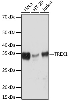
Western blot analysis of various lysates using TREX1 Rabbit mAb (CAB3819) at 1:1000 dilution. Secondary antibody: HRP-conjugated Goat anti-Rabbit IgG (H+L) (CABS014) at 1:10000 dilution. Lysates/proteins: 25μg per lane. Blocking buffer: 3% nonfat dry milk in TBST. Detection: ECL Basic Kit (AbGn00020). Exposure time: 10s.
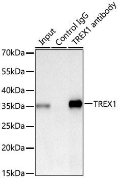
Immunoprecipitation of TREX1 from 300 µg extracts of HeLa cells was performed using 0.5 µg of TREX1 Rabbit mAb (CAB3819). Rabbit IgG isotype control (AC005) was used to precipitate the Control IgG sample. IP samples were eluted with 1X reducing Laemmli Buffer. The Input lane represents 10% of the total input. Western blot analysis of immunoprecipitates was conducted using TREX1 Rabbit mAb (CAB3819) at a dilution of 1:1000.