The TRH Antibody (CAB8322) is a high-quality antibody developed for reliable detection and analysis of target proteins. This antibody, produced in rabbits, is highly specific for TRH and has been validated for use in various applications including immunohistochemistry, ELISA, and Western blotting.TRH, also known as thyrotropin-releasing hormone, plays a crucial role in the hypothalamic-pituitary-thyroid axis, regulating the release of thyroid-stimulating hormone (TSH) and ultimately affecting thyroid hormone production.
This antibody is validated for use in WB, ELISA, IF-P applications and has demonstrated reactivity against Human, Mouse, Rat samples.
Product Name:
TRH Antibody
SKU:
CAB8322
Size:
20μL, 100μL
Reactivity:
Human, Mouse, Rat
Conjugate:
Unconjugated
Immunogen:
Synthetic peptide. This information is considered to be commercially sensitive.
Recommended starting concentration is 1 μg/mL. Please optimize the concentration based on your specific assay requirements.
Synonyms:
TRF, Pro-TRH, TRH
Positive Sample:
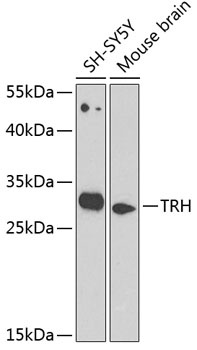
SH-SY5Y, Mouse brain
Cellular Localization:
Secreted.
Calculated MW:
27kDa
Observed MW:
27kDa
This gene encodes a member of the thyrotropin-releasing hormone family. Cleavage of the encoded proprotein releases mature thyrotropin-releasing hormone, which is a tripeptide hypothalamic regulatory hormone. The human proprotein contains six thyrotropin-releasing hormone tripeptides. Thyrotropin-releasing hormone is involved in the regulation and release of thyroid-stimulating hormone, as well as prolactin. Deficiency of this hormone has been associated with hypothalamic hypothyroidism.
Purification Method
Affinity purification
Gene ID
7200
RRID
AB_2772703
Buffer Information
Store at -20℃. Avoid freeze / thaw cycles. Buffer: PBS containing 50% glycerol, preserved with proclin300 or sodium azide, pH 7.3.
Western blot analysis of various lysates using TRH Rabbit pAb (CAB8322) at 1:1000 dilution. Secondary antibody: HRP-conjugated Goat anti-Rabbit IgG (H+L) (CABS014) at 1:10000 dilution. Lysates/proteins: 25μg per lane. Blocking buffer: 3% nonfat dry milk in TBST. Detection: ECL Basic Kit (AbGn00020). Exposure time: 90s.
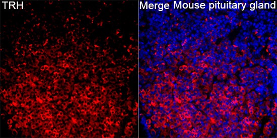
Immunofluorescence analysis of Mouse pituitary gland tissue using TRH Rabbit pAb (CAB8322) at a dilution of 1:300 (40x lens). Secondary antibody: Cy3 Goat Anti-Rabbit IgG (H+L)(CABS007) at 1:500 dilution. Blue: DAPI for nuclear staining. High pressure antigen retrieval performed with 0.01M Citrate Buffer (pH 6.0) prior to IF staining.