The TRITC-conjugated Goat anti-Mouse IgG (H+L) (CABS026) is a high-quality antibody developed for reliable detection and analysis of target proteins. This antibody, raised in goats and conjugated with TRITC for fluorescent labeling, is highly specific and sensitive for detecting mouse IgG in samples. It is ideal for use in immunofluorescence, flow cytometry, and other fluorescence-based assays.The TRITC-conjugated Goat anti-Mouse IgG (H+L) antibody binds specifically to mouse IgG, allowing for easy and efficient detection and analysis of mouse IgG in various experimental settings.
This antibody is validated for use in IF/ICC, FC applications and has demonstrated reactivity against Mouse samples.
Product Name:
TRITC-conjugated Goat anti-Mouse IgG (H+L)
SKU:
CABS026
Size:
100μL
Reactivity:
Mouse
Conjugate:
Rhodamine. Ex:550nm. Em:570nm.
Immunogen:
This information is considered to be commercially sensitive.
Tested Applications:
IF/ICCFC
Recommended Dilution:
IF/ICC
1:50 - 1:200
FC
1:50 - 1:200
Secondary antibodies are affinity-purified antibodies which will work with target-specific primary antibody in the detection, sorting or purification of its specified target. Secondary antibodies offer increased versatility enabling users to use many detection systems (e.g. HRP, AP, fluorescence). They can also provide greater sensitivity through signal amplification as multiple secondary antibodies . Most commonly, secondary antibodies are generated by immunizing the host animal (different from host species of primary antibody) with a pooled population of normal immunoglobulins from the host species of primary antibody and can be further purified and modified (i.e. antibody fragmentation, label conjugation, etc.) to ensure well-characterized specificity to corresponding normal immunoglobulins.
Purification Method
Affinity purification
RRID
AB_2772721
Buffer Information
Store at -20℃. Avoid freeze / thaw cycles. Buffer: PBS with 0.025% Sodium Azide,0.75% BSA,50% glycerol,pH7.3.
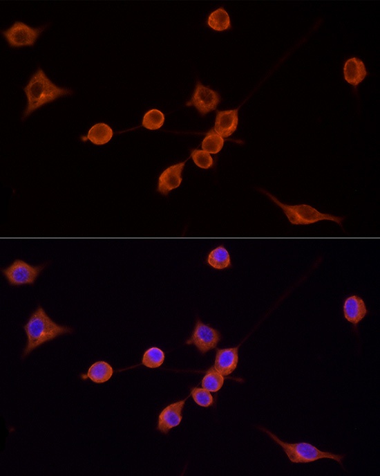
Immunofluorescence analysis of NIH/3T3 cells using TRITC Goat Anti-Mouse IgG (H+L) (CABS026) at dilution of 1:200 (40x lens). Blue: DAPI for nuclear staining.
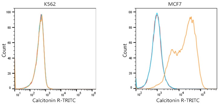
Flow cytometric analysis of Positive antibody Human Calcitonin R (2.5μg/mL) in various cells (orange) compare to Mouse isotype control (blue) and non-staining control (Red). The secondary antibody used was TRITC Goat Anti-Mouse IgG (H+L) (CABS026) at 1:100.
 (CABS026)")
 (CABS026)")
-conjugated Goat anti-Rabbit IgG (H+L) (CABS040)")
 (CABS008)")
 (CABS017)")
 (CABS001)")
 (CABS003)")