The TRPA1 Antibody (CAB8568) is a high-quality antibody developed for reliable detection and analysis of target proteins. This antibody, produced in rabbits, is highly specific for human samples and has been validated for use in Western blot applications. By targeting the TRPA1 protein, this antibody allows for precise detection and analysis in a variety of cell types, making it an ideal choice for studies in neuroscience, pain research, and drug development.
This antibody is validated for use in WB, IHC-P, ELISA, IF-P applications and has demonstrated reactivity against Human, Mouse, Rat samples.
Product Name:
TRPA1 Antibody
SKU:
CAB8568
Size:
20μL, 100μL
Reactivity:
Human, Mouse, Rat
Conjugate:
Unconjugated
Immunogen:
Synthetic peptide. This information is considered to be commercially sensitive.
Recommended starting concentration is 1 μg/mL. Please optimize the concentration based on your specific assay requirements.
Synonyms:
FEPS, p120, FEPS1, ANKTM1, TRPA1
Positive Sample:
Mouse brain
Cellular Localization:
Cell Membrane, Multi-Pass Membrane Protein.
Calculated MW:
128kDa
Observed MW:
128kDa
The structure of the protein encoded by this gene is highly related to both the protein ankyrin and transmembrane proteins. The specific function of this protein has not yet been determined; however, studies indicate the function may involve a role in signal transduction and growth control.
Purification Method
Affinity purification
Gene ID
8989
RRID
AB_2772726
Buffer Information
Store at -20℃. Avoid freeze / thaw cycles. Buffer: PBS containing 50% glycerol, preserved with proclin300 or sodium azide, pH 7.3.
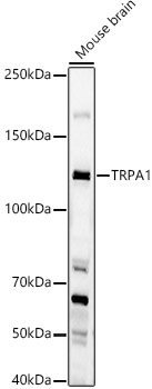
Western blot analysis of lysates from Mouse brain, using TRPA1 Rabbit pAb (CAB8568) at 1:1000 dilution. Secondary antibody: HRP-conjugated Goat anti-Rabbit IgG (H+L) (CABS014) at 1:10000 dilution. Lysates/proteins: 25μg per lane. Blocking buffer: 3% nonfat dry milk in TBST. Detection: ECL Basic Kit (AbGn00020). Exposure time: 60s.
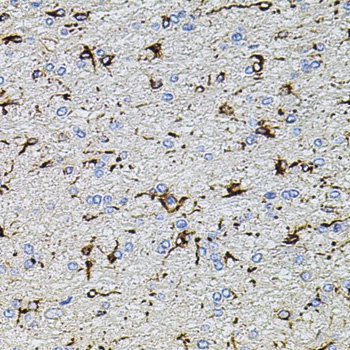
Immunohistochemistry analysis of paraffin-embedded Rat brain using TRPA1 Rabbit pAb (CAB8568) at dilution of 1:100 (40x lens). Microwave antigen retrieval performed with 0.01M PBS Buffer (pH 7.2) prior to IHC staining.
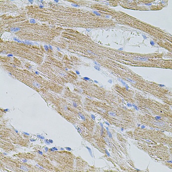
Immunohistochemistry analysis of paraffin-embedded Mouse heart using TRPA1 Rabbit pAb (CAB8568) at dilution of 1:100 (40x lens). Microwave antigen retrieval performed with 0.01M PBS Buffer (pH 7.2) prior to IHC staining.
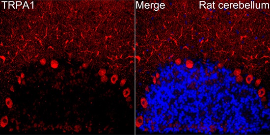
Immunofluorescence analysis of paraffin-embedded Rat cerebellum tissue using TRPA1 Rabbit pAb(CAB8568) at a dilution of 1:200 (40x lens). Secondary antibody:Cy3 Goat Anti-Rabbit IgG (H+L)(CABS007) at 1:500 dilution. Blue: DAPI for nuclear staining. Perform microwave antigen retrieval with 0.01 M citrate buffer (pH 6.0) prior to IF staining.