The TSFM Antibody (CAB16094) is a high-quality antibody developed for reliable detection and analysis of target proteins. This antibody, produced in rabbits, offers high reactivity with human samples and has been validated for use in Western blotting and immunohistochemistry applications.TSFM is a key component in the translation machinery of mitochondria, playing a crucial role in protein synthesis within these energy-producing organelles. Dysregulation of TSFM has been associated with various mitochondrial disorders and diseases, making it a target of interest in research on metabolic disorders, neurodegenerative diseases, and cancer.
This antibody is validated for use in WB, IF/ICC, ELISA applications and has demonstrated reactivity against Human, Mouse, Rat samples.
Product Name:
TSFM Antibody
SKU:
CAB16094
Size:
20μL, 100μL
Reactivity:
Human, Mouse, Rat
Conjugate:
Unconjugated
Immunogen:
Recombinant protein (or fragment).This information is considered to be commercially sensitive.
Recommended starting concentration is 1 μg/mL. Please optimize the concentration based on your specific assay requirements.
Synonyms:
EFTS, EFTSMT, TSFM
Positive Sample:
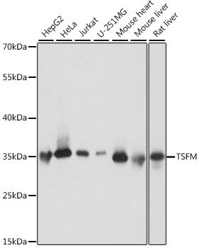
HepG2, HeLa, Jurkat, U-251MG, Mouse heart, Mouse liver, Rat liver
Cellular Localization:
Mitochondrion.
Calculated MW:
35kDa
Observed MW:
35kDa
This gene encodes a mitochondrial translation elongation factor. The encoded protein is an enzyme that catalyzes the exchange of guanine nucleotides on the translation elongation factor Tu during the elongation step of mitchondrial protein translation. Mutations in this gene are associated with combined oxidative phosphorylation deficiency-3 syndrome. Alternate splicing results in multiple transcript variants.
Purification Method
Affinity purification
Gene ID
10102
RRID
AB_2763536
Buffer Information
Store at -20℃. Avoid freeze / thaw cycles. Buffer: PBS with 0.01% thimerosal,50% glycerol,pH7.3.
Western blot analysis of various lysates using TSFM Rabbit pAb (CAB16094) at 1:1000 dilution. Secondary antibody: HRP-conjugated Goat anti-Rabbit IgG (H+L) (CABS014) at 1:10000 dilution. Lysates/proteins: 25μg per lane. Blocking buffer: 3% nonfat dry milk in TBST. Detection: ECL Basic Kit (AbGn00020). Exposure time: 1s.
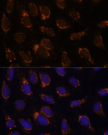
Immunofluorescence analysis of U-2 OS cells using TSFM Rabbit pAb (CAB16094) at dilution of 1:100 (40x lens). Secondary antibody: Cy3-conjugated Goat anti-Rabbit IgG (H+L) (CABS007) at 1:500 dilution. Blue: DAPI for nuclear staining.