The TSHR Antibody (CAB6781) is a high-quality antibody developed for reliable detection and analysis of target proteins. The protein encoded by this gene is a membrane protein and a major controller of thyroid cell metabolism. The encoded protein is a receptor for thyrothropin and thyrostimulin, and its activity is mediated by adenylate cyclase. Defects in this gene are a cause of several types of hyperthyroidism. Three transcript variants encoding different isoforms have been found for this gene.
This antibody is validated for use in WB, IHC-P, ELISA applications and has demonstrated reactivity against Human, Mouse, Rat samples.
Product Name:
TSHR Antibody
SKU:
CAB6781
Size:
100μL, 20μL
Reactivity:
Human, Mouse, Rat
Conjugate:
Unconjugated
Immunogen:
Recombinant protein (or fragment).This information is considered to be commercially sensitive.
Tested Applications:
WBIHC-PELISA
Recommended Dilution:
WB
1:500 - 1:1000
IHC-P
1:50 - 1:200
ELISA
Recommended starting concentration is 1 μg/mL. Please optimize the concentration based on your specific assay requirements.
Synonyms:
LGR3, CHNG1, hTSHR-I, TSHR
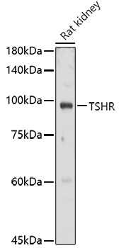
Positive Sample:
Rat kidney
Cellular Localization:
Cell Membrane, Multi-Pass Membrane Protein.
Calculated MW:
87kDa
Observed MW:
100kDa
The protein encoded by this gene is a membrane protein and a major controller of thyroid cell metabolism. The encoded protein is a receptor for thyrothropin and thyrostimulin, and its activity is mediated by adenylate cyclase. Defects in this gene are a cause of several types of hyperthyroidism. Three transcript variants encoding different isoforms have been found for this gene.
Purification Method
Affinity purification
Gene ID
7253
RRID
AB_2767364
Buffer Information
Store at -20℃. Avoid freeze / thaw cycles. Buffer: PBS containing 50% glycerol, preserved with proclin300 or sodium azide, pH 7.3.
Western blot analysis of lysates from Rat kidney, using TSHR Rabbit pAb (CAB6781) at 1:1000 dilution. Secondary antibody: HRP-conjugated Goat anti-Rabbit IgG (H+L) (AS014) at 1:10000 dilution. Lysates/proteins: 25μg per lane. Blocking buffer: 3% nonfat dry milk in TBST. Detection: ECL Basic Kit (AbGn00020). Exposure time: 30s.
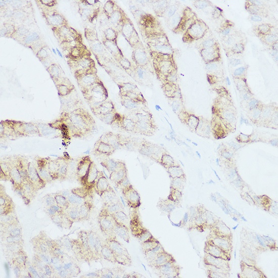
Immunohistochemistry analysis of paraffin-embedded Human thyroid cancer using TSHR Rabbit pAb (CAB6781) at dilution of 1:200 (40x lens). High pressure antigen retrieval performed with 0.01M Citrate buffer (pH 6.0) prior to IHC staining.