The β-Tubulin Rabbit pAb (CABC015) is a high-quality antibody developed for reliable detection and analysis of target proteins. This antibody, generated from rabbit immunizations, exhibits high reactivity with tubulin in human samples and is suitable for use in Western blot applications. By specifically binding to tubulin, this antibody enables precise detection and analysis in a variety of cell types, making it an essential reagent for studies in cell biology, cancer research, and drug development.Tubulin plays a critical role in maintaining cell shape and structure, as well as facilitating crucial processes such as mitosis and intracellular trafficking.
This antibody is validated for use in WB, IHC-P, IF/ICC applications and has demonstrated reactivity against Human, Mouse, Rat samples.
Product Name:
β-Tubulin Rabbit pAb
SKU:
CABC015
Size:
100μL
Reactivity:
Human, Mouse, Rat
Conjugate:
Unconjugated
Immunogen:
Recombinant protein (or fragment).This information is considered to be commercially sensitive.
SH-SY5Y, HeLa, A-549, MCF7, HepG2, Mouse eye, Rat spinal cord
Cellular Localization:
Cytoplasm, Cytoskeleton.
Calculated MW:
50kDa
Observed MW:
55kDa
This gene encodes a beta tubulin protein. This protein forms a dimer with alpha tubulin and acts as a structural component of microtubules. Mutations in this gene cause cortical dysplasia, complex, with other brain malformations 6. Alternative splicing results in multiple splice variants. There are multiple pseudogenes for this gene on chromosomes 1, 6, 7, 8, 9, and 13.
Purification Method
Affinity purification
Gene ID
203068
RRID
AB_2773007
Buffer Information
Store at -20℃. Avoid freeze / thaw cycles. Buffer: PBS with 0.09% Sodium azide,50% glycerol,pH7.3.
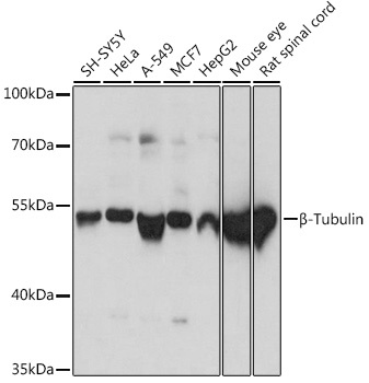
Western blot analysis of various lysates using β-Tubulin Rabbit pAb (CABC015) at 1:1000 dilution. Secondary antibody: HRP-conjugated Goat anti-Rabbit IgG (H+L) (CABS014) at 1:10000 dilution. Lysates/proteins: 25μg per lane. Blocking buffer: 3% nonfat dry milk in TBST. Detection: ECL Basic Kit (AbGn00020). Exposure time: 3s.
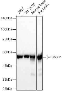
Western blot analysis of various lysates, using β-Tubulin Rabbit pAb (CABC015) at 1:2000 dilution. Secondary antibody: HRP-conjugated Goat anti-Rabbit IgG (H+L) (CABS014) at 1:10000 dilution. Lysates/proteins: 25μg per lane. Blocking buffer: 3% nonfat dry milk in TBST. Detection: ECL Basic Kit (AbGn00020). Exposure time: 1s.
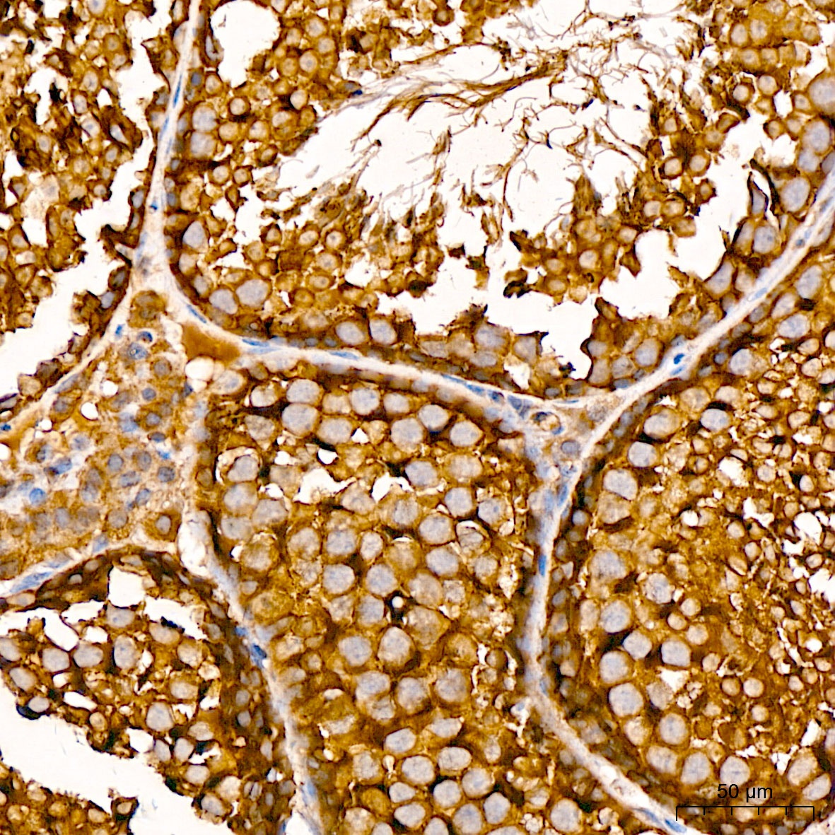
Immunohistochemistry analysis of paraffin-embedded Mouse testis tissue using β-Tubulin Rabbit pAb (CABC015) at a dilution of 1:100 (40x lens). High pressure antigen retrieval was performed with 0.01 M citrate buffer (pH 6.0) prior to IHC staining.
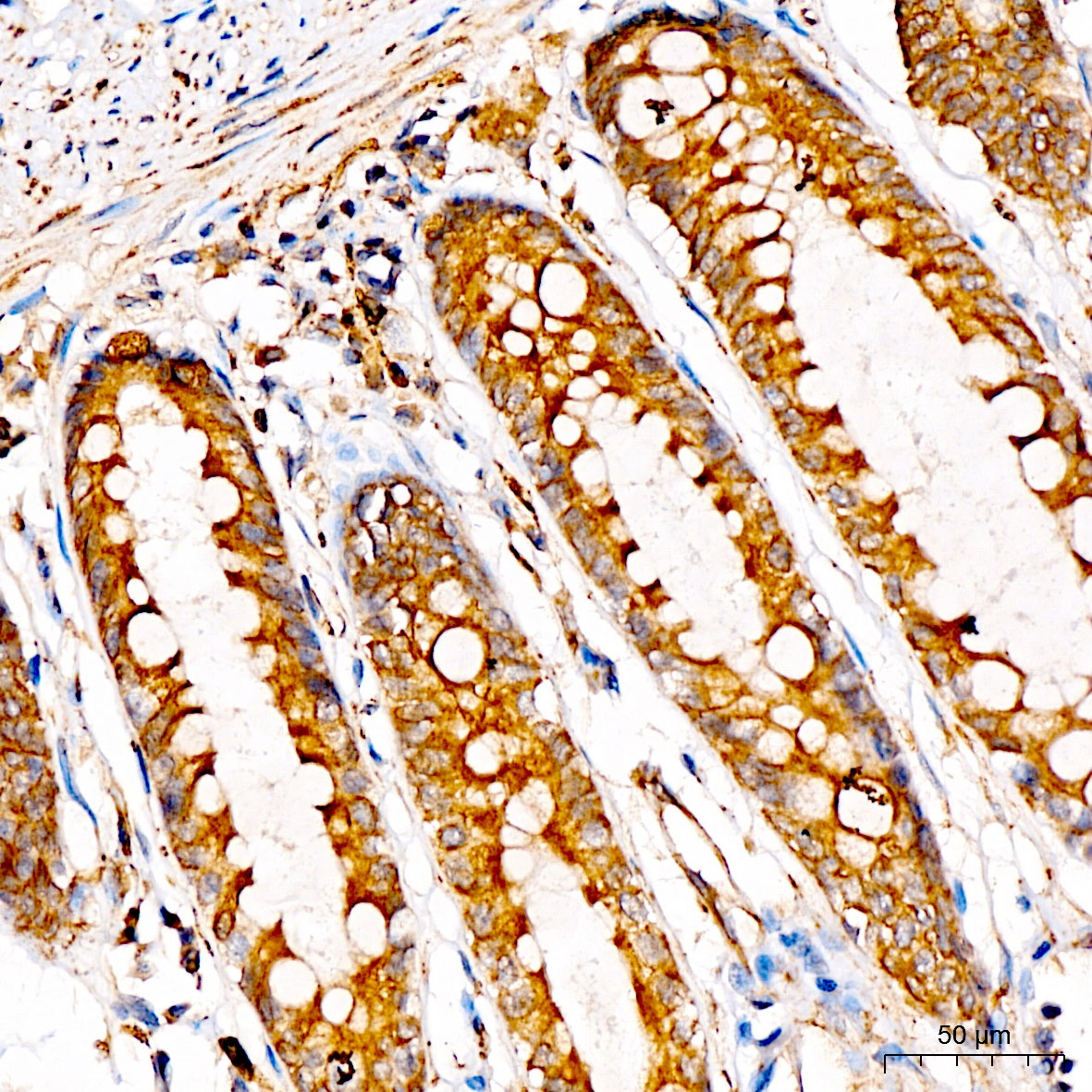
Immunohistochemistry analysis of paraffin-embedded Human colon tissue using β-Tubulin Rabbit pAb (CABC015) at a dilution of 1:100 (40x lens). High pressure antigen retrieval was performed with 0.01 M citrate buffer (pH 6.0) prior to IHC staining.
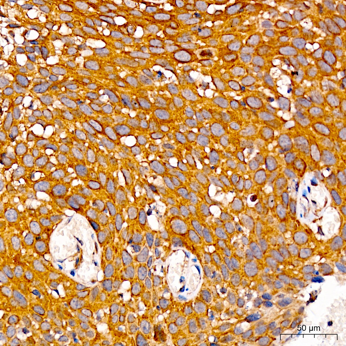
Immunohistochemistry analysis of paraffin-embedded Human cervix cancer tissue using β-Tubulin Rabbit pAb (CABC015) at a dilution of 1:100 (40x lens). High pressure antigen retrieval was performed with 0.01 M citrate buffer (pH 6.0) prior to IHC staining.
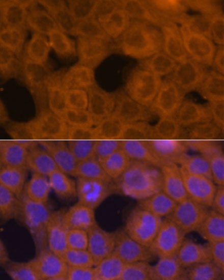
Immunofluorescence analysis of A431 cells using β-Tubulin Rabbit pAb (CABC015) at dilution of 1:100. Secondary antibody: Cy3-conjugated Goat anti-Rabbit IgG (H+L) (CABS007) at 1:500 dilution. Blue: DAPI for nuclear staining.
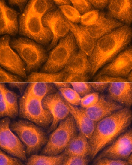
Immunofluorescence analysis of HeLa cells using β-Tubulin Rabbit pAb (CABC015) at dilution of 1:100. Secondary antibody: Cy3-conjugated Goat anti-Rabbit IgG (H+L) (CABS007) at 1:500 dilution. Blue: DAPI for nuclear staining.
")
")
")
")
")
")
")