The Tulp2 Polyclonal Antibody (CAB18256) is a high-quality antibody developed for reliable detection and analysis of target proteins. This antibody, produced in rabbits, is highly specific to human samples and is validated for use in Western blot applications. By targeting the TULP2 protein, this antibody enables researchers to detect and analyze TULP2 expression in a variety of cell types, making it an essential tool for studies in cell biology and neuroscience.TULP2, also known as tubby like protein 2, is known to play important roles in neuronal development, ciliary trafficking, and cell signaling. Its involvement in these processes highlights its relevance in diseases such as retinal degeneration, obesity, and neurological disorders.
This antibody is validated for use in WB, IHC-P, ELISA applications and has demonstrated reactivity against Human, Mouse, Rat samples.
Product Name:
Tulp2 Polyclonal Antibody
SKU:
CAB18256
Size:
20μL, 100μL
Reactivity:
Human, Mouse, Rat
Conjugate:
Unconjugated
Immunogen:
Recombinant protein (or fragment).This information is considered to be commercially sensitive.
Recommended starting concentration is 1 μg/mL. Please optimize the concentration based on your specific assay requirements.
Synonyms:
Pdet, Tulp2
Positive Sample:
NIH/3T3, PC-3, SKOV3, Rat testis
Cellular Localization:
Cilium, Cytoplasm, Extracellular Region.
Calculated MW:
63kDa
Observed MW:
60kDa
Predicted to enable phosphoric diester hydrolase activity. Predicted to be involved in protein localization to cilium. Predicted to be located in cytoplasm and extracellular region. Predicted to be active in cilium. Is expressed in urogenital ridge. Orthologous to human TULP2 (TUB like protein 2).
Purification Method
Affinity purification
Gene ID
56734
RRID
AB_2862032
Buffer Information
Store at -20℃. Avoid freeze / thaw cycles. Buffer: PBS containing 50% glycerol, preserved with proclin300 or sodium azide, pH 7.3.
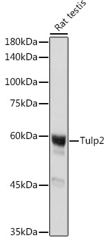
Western blot analysis of lysates from Rat testis, using Tulp2 Rabbit pAb (CAB18256) at 1:1000 dilution. Secondary antibody: HRP-conjugated Goat anti-Rabbit IgG (H+L) (CABS014) at 1:10000 dilution. Lysates/proteins: 25μg per lane. Blocking buffer: 3% nonfat dry milk in TBST. Detection: ECL Basic Kit (AbGn00020). Exposure time: 1s.
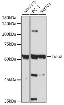
Western blot analysis of various lysates using Tulp2 Rabbit pAb (CAB18256) at 1:1000 dilution. Secondary antibody: HRP-conjugated Goat anti-Rabbit IgG (H+L) (CABS014) at 1:10000 dilution. Lysates/proteins: 25μg per lane. Blocking buffer: 3% nonfat dry milk in TBST. Detection: ECL Enhanced Kit (AbGn00021). Exposure time: 30s.
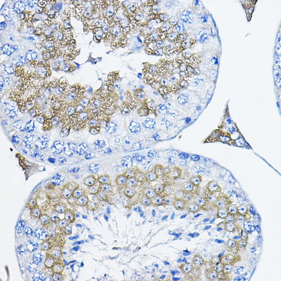
Immunohistochemistry analysis of paraffin-embedded Mouse testis using Tulp2 Rabbit pAb (CAB18256) at dilution of 1:200 (40x lens). High pressure antigen retrieval performed with 0.01M Citrate buffer (pH 6.0) prior to IHC staining.