The p63 Antibody (CAB2137) is a high-quality antibody developed for reliable detection and analysis of target proteins. This antibody, generated in rabbits, exhibits high reactivity with human samples and is validated for use in Western blot applications.TP63, a member of the p53 tumor suppressor gene family, plays a critical role in various cellular processes, including cell cycle regulation, apoptosis, and differentiation. Dysregulation of TP63 has been implicated in a wide range of cancers, making it a promising target for cancer research and therapy development.
This antibody is validated for use in WB, IHC-P, ELISA, IF-P applications and has demonstrated reactivity against Human, Mouse, Rat samples.
Product Name:
p63 Antibody
SKU:
CAB2137
Size:
20μL, 100μL
Reactivity:
Human, Mouse, Rat
Conjugate:
Unconjugated
Immunogen:
Synthetic peptide. This information is considered to be commercially sensitive.
This gene encodes a member of the p53 family of transcription factors. The functional domains of p53 family proteins include an N-terminal transactivation domain, a central DNA-binding domain and an oligomerization domain. Alternative splicing of this gene and the use of alternative promoters results in multiple transcript variants encoding different isoforms that vary in their functional properties. These isoforms function during skin development and maintenance, adult stem/progenitor cell regulation, heart development and premature aging. Some isoforms have been found to protect the germline by eliminating oocytes or testicular germ cells that have suffered DNA damage. Mutations in this gene are associated with ectodermal dysplasia, and cleft lip/palate syndrome 3 (EEC3); split-hand/foot malformation 4 (SHFM4); ankyloblepharon-ectodermal defects-cleft lip/palate; ADULT syndrome (acro-dermato-ungual-lacrimal-tooth); limb-mammary syndrome; Rap-Hodgkin syndrome (RHS); and orofacial cleft 8.
Purification Method
Affinity purification
Gene ID
8626
RRID
AB_2764156
Buffer Information
Store at -20℃. Avoid freeze / thaw cycles. Buffer: PBS with 0.09% Sodium azide,50% glycerol,pH7.3.
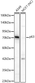
Western blot analysis of various lysates, using p63 Rabbit pAb (CAB2137) at 1:1000 dilution. Secondary antibody: HRP-conjugated Goat anti-Rabbit IgG (H+L) (CABS014) at 1:10000 dilution. Lysates/proteins: 25μg per lane. Blocking buffer: 3% nonfat dry milk in TBST. Detection: ECL Basic Kit (AbGn00020). Negative control (NC):MCF7 Exposure time: 10s.
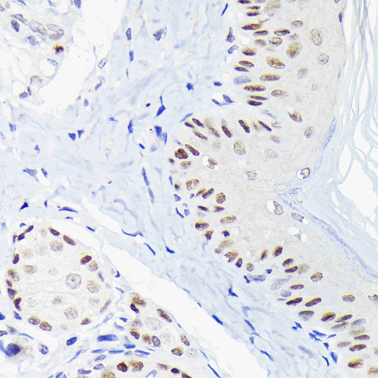
Immunohistochemistry analysis of paraffin-embedded Rat skin using p63 Rabbit pAb (CAB2137) at dilution of 1:100 (40x lens). Microwave antigen retrieval performed with 0.01M Tris/EDTA Buffer (pH 9.0) prior to IHC staining.
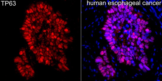
Immunofluorescence analysis of human esophageal cancer using p63 Rabbit pAb (CAB2137) at dilution of 1:100 (40x lens). Secondary antibody: Cy3-conjugated Goat anti-Rabbit IgG (H+L) (CABS007) at 1:500 dilution. Blue: DAPI for nuclear staining.