The TXLNA Antibody (CAB18291) is a high-quality antibody developed for reliable detection and analysis of target proteins. This antibody, produced in rabbits, exhibits high reactivity with human samples and has been validated for use in Western blot applications.TXLNA, also known as transcription elongation factor A (SII)-like 3, is essential for maintaining genomic integrity and promoting proper gene expression. Dysregulation of TXLNA has been linked to diseases such as cancer and neurodegenerative disorders, making it a promising target for therapeutic interventions.
This antibody is validated for use in WB, IF/ICC, ELISA applications and has demonstrated reactivity against Human samples.
Product Name:
TXLNA Antibody
SKU:
CAB18291
Size:
20μL, 100μL
Reactivity:
Human
Immunogen:
Recombinant protein (or fragment).This information is considered to be commercially sensitive.
Recommended starting concentration is 1 μg/mL. Please optimize the concentration based on your specific assay requirements.
Synonyms:
IL14, TXLN, TXLNA
Positive Sample:
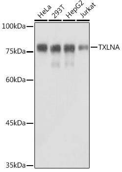
HeLa, 293T, HepG2, Jurkat
Cellular Localization:
Cytoplasm, Cytosol, Extracellular Region.
Calculated MW:
62kDa
Observed MW:
75kDa
Predicted to enable syntaxin binding activity. Predicted to be involved in exocytosis. Predicted to act upstream of or within B cell activation. Located in cytoplasm.
Purification Method
Affinity purification
Gene ID
200081
RRID
AB_2862064
Buffer Information
Store at -20℃. Avoid freeze / thaw cycles. Buffer: PBS containing 50% glycerol, preserved with proclin300 or sodium azide, pH 7.3.
Western blot analysis of various lysates using TXLNA Rabbit pAb (CAB18291) at 1:1000 dilution. Secondary antibody: HRP-conjugated Goat anti-Rabbit IgG (H+L) (CABS014) at 1:10000 dilution. Lysates/proteins: 25μg per lane. Blocking buffer: 3% nonfat dry milk in TBST. Detection: ECL Basic Kit (AbGn00020). Exposure time: 1s.
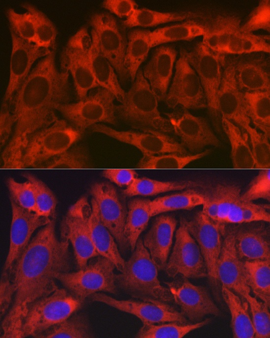
Immunofluorescence analysis of U2OS cells using TXLNA Rabbit pAb (CAB18291) at dilution of 1:100 (40x lens). Secondary antibody: Cy3-conjugated Goat anti-Rabbit IgG (H+L) (CABS007) at 1:500 dilution. Blue: DAPI for nuclear staining.


")


