The TXNDC5 Antibody (CAB14152) is a high-quality antibody developed for reliable detection and analysis of target proteins. This antibody, produced in rabbits, is highly specific for detecting TXNDC5 in human samples and has been validated for use in Western blot applications. By binding to TXNDC5, this antibody allows for the visualization and analysis of the protein in various cell types, making it ideal for studies in oxidative stress, cellular signaling, and disease mechanisms.TXNDC5, also known as thioredoxin domain-containing protein 5, plays a critical role in maintaining cellular redox balance and protecting cells from oxidative damage. Dysregulation of TXNDC5 has been implicated in various diseases, including cancer, neurodegenerative disorders, and cardiovascular disease.
This antibody is validated for use in WB, IHC-P, IF/ICC, ELISA applications and has demonstrated reactivity against Human, Mouse, Rat samples.
Product Name:
TXNDC5 Antibody
SKU:
CAB14152
Size:
20μL, 100μL
Reactivity:
Human, Mouse, Rat
Conjugate:
Unconjugated
Immunogen:
Recombinant protein (or fragment).This information is considered to be commercially sensitive.
This gene encodes a member of the disulfide isomerase (PDI) family of endoplasmic reticulum (ER) proteins that catalyze protein folding and thiol-disulfide interchange reactions. The encoded protein has an N-terminal endoplasmic reticulum (ER)-signal sequence, three catalytically active thioredoxin domains and a C-terminal ER-retention sequence. Its expression is induced by hypoxia and its role may be to protect hypoxic cells from apoptosis. Alternative splicing results in multiple transcript variants. Read-through transcription also exists between this gene and the neighboring upstream BLOC1S5 gene.
Purification Method
Affinity purification
Gene ID
81567
RRID
AB_2761010
Buffer Information
Store at -20℃. Avoid freeze / thaw cycles. Buffer: PBS containing 50% glycerol, preserved with proclin300 or sodium azide, pH 7.3.
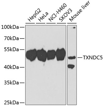
Western blot analysis of various lysates using TXNDC5 Rabbit pAb (CAB14152) at 1:1000 dilution. Secondary antibody: HRP-conjugated Goat anti-Rabbit IgG (H+L) (CABS014) at 1:10000 dilution. Lysates/proteins: 25μg per lane. Blocking buffer: 3% nonfat dry milk in TBST. Detection: ECL Basic Kit (AbGn00020). Exposure time: 90s.
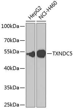
Western blot analysis of various lysates using TXNDC5 Rabbit pAb (CAB14152) at 1:1000 dilution. Secondary antibody: HRP-conjugated Goat anti-Rabbit IgG (H+L) (CABS014) at 1:10000 dilution. Lysates/proteins: 25μg per lane. Blocking buffer: 3% nonfat dry milk in TBST. Detection: ECL Basic Kit (AbGn00020). Exposure time: 90s.
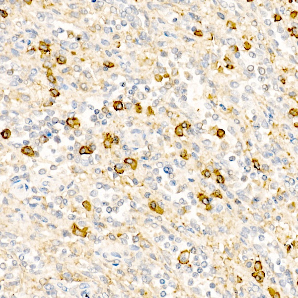
Immunohistochemistry analysis of paraffin-embedded Human spleen tissue using TXNDC5 Rabbit pAb (CAB14152) at a dilution of 1:500 (40x lens). High pressure antigen retrieval was performed with 0.01 M citrate buffer (pH 6.0) prior to IHC staining.
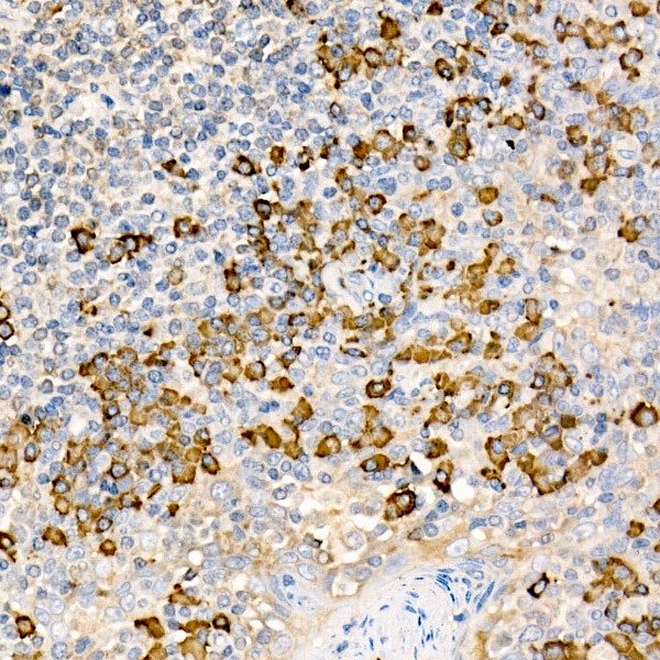
Immunohistochemistry analysis of paraffin-embedded Human tonsil tissue using TXNDC5 Rabbit pAb (CAB14152) at a dilution of 1:500 (40x lens). High pressure antigen retrieval was performed with 0.01 M citrate buffer (pH 6.0) prior to IHC staining.
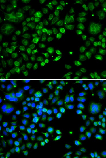
Immunofluorescence analysis of MCF-7 cells using TXNDC5 Rabbit pAb (CAB14152). Secondary antibody: Cy3-conjugated Goat anti-Rabbit IgG (H+L) (CABS007) at 1:500 dilution. Blue: DAPI for nuclear staining.