The TXNIP Polyclonal Antibody (CAB24289) is a high-quality antibody developed for reliable detection and analysis of target proteins. This antibody, generated in rabbits, exhibits high specificity and sensitivity towards human samples, making it ideal for use in Western blot applications.TXNIP, also known as thioredoxin-interacting protein, is a key regulator of redox balance in cells, playing a crucial role in mediating oxidative stress responses and controlling glucose uptake. Dysregulation of TXNIP has been implicated in various diseases, including diabetes, cardiovascular disorders, and cancer.
This antibody is validated for use in WB, ELISA applications and has demonstrated reactivity against Mouse, Rat samples.
Product Name:
TXNIP Polyclonal Antibody
SKU:
CAB24289
Size:
20μL, 100μL
Reactivity:
Mouse, Rat
Conjugate:
Unconjugated
Immunogen:
Synthetic peptide. This information is considered to be commercially sensitive.
Recommended starting concentration is 1 μg/mL. Please optimize the concentration based on your specific assay requirements.
Synonyms:
THIF, VDUP1, ARRDC6, HHCPA78, EST01027, TXNIP
Positive Sample:
Mouse thymus
Cellular Localization:
Cytoplasm.
Calculated MW:
44kDa
Observed MW:
55kDa
This gene encodes a thioredoxin-binding protein that is a member of the alpha arrestin protein family. Thioredoxin is a thiol-oxidoreductase that is a major regulator of cellular redox signaling which protects cells from oxidative stress. This protein inhibits the antioxidative function of thioredoxin resulting in the accumulation of reactive oxygen species and cellular stress. This protein also functions as a regulator of cellular metabolism and of endoplasmic reticulum (ER) stress. This protein may also function as a tumor suppressor. Alternate splicing results in multiple transcript variants.
Purification Method
Affinity purification
Gene ID
10628
Buffer Information
Store at -20℃. Avoid freeze / thaw cycles. Buffer: PBS containing 50% glycerol, preserved with proclin300 or sodium azide, pH 7.3.
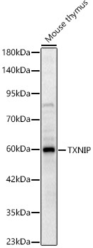
Western blot analysis of lysates from Mouse thymus, using TXNIP Rabbit pAb (CAB24289) at 1:2000 dilution. Secondary antibody: HRP-conjugated Goat anti-Rabbit IgG (H+L) (CABS014) at 1:10000 dilution. Lysates/proteins: 25μg per lane. Blocking buffer: 3% nonfat dry milk in TBST. Detection: ECL Basic Kit (AbGn00020). Exposure time: 60s.
 at 1:2000 dilution. Secondary antibody: HRP Goat Anti-Rabbit IgG (H+L) at 1:10000 dilution. Lysates/proteins: 25ug per lane. Blocking buffer: 3% nonfat dry milk in TBST.")
 at 1:2000 dilution. Secondary antibody: HRP Goat Anti-Rabbit IgG (H+L) at 1:10000 dilution. Lysates/proteins: 25ug per lane. Blocking buffer: 3% nonfat dry milk in TBST.")


