The TXNIP Antibody (CAB9342) is a high-quality antibody developed for reliable detection and analysis of target proteins. This rabbit-derived antibody offers highly specific and sensitive detection of TXNIP in human samples, making it ideal for Western blot applications. By targeting the TXNIP protein, researchers can gain insights into its role in regulating glucose metabolism, apoptosis, and oxidative stress pathways in various cell types.TXNIP, also known as Vitamin D3 up-regulated protein 1 (VDUP1), is involved in multiple cellular processes, including glucose uptake and insulin sensitivity, making it a potential therapeutic target for metabolic disorders such as diabetes and obesity.
This antibody is validated for use in WB, IF/ICC, ELISA applications and has demonstrated reactivity against Human, Mouse, Rat samples.
Product Name:
TXNIP Antibody
SKU:
CAB9342
Size:
20μL, 100μL
Reactivity:
Human, Mouse, Rat
Conjugate:
Unconjugated
Immunogen:
Synthetic peptide. This information is considered to be commercially sensitive.
Recommended starting concentration is 1 μg/mL. Please optimize the concentration based on your specific assay requirements.
Synonyms:
THIF, VDUP1, ARRDC6, HHCPA78, EST01027, TXNIP
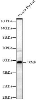
Positive Sample:
Mouse thymus
Cellular Localization:
Cytoplasm.
Calculated MW:
44kDa
Observed MW:
55kDa
This gene encodes a thioredoxin-binding protein that is a member of the alpha arrestin protein family. Thioredoxin is a thiol-oxidoreductase that is a major regulator of cellular redox signaling which protects cells from oxidative stress. This protein inhibits the antioxidative function of thioredoxin resulting in the accumulation of reactive oxygen species and cellular stress. This protein also functions as a regulator of cellular metabolism and of endoplasmic reticulum (ER) stress. This protein may also function as a tumor suppressor. Alternate splicing results in multiple transcript variants.
Purification Method
Affinity purification
Gene ID
10628
RRID
AB_2772765
Buffer Information
Store at -20℃. Avoid freeze / thaw cycles. Buffer: PBS with 0.09% Sodium azide,50% glycerol,pH7.3.
Western blot analysis of lysates from Mouse thymus, using TXNIP Rabbit pAb (CAB9342) at 1:2000 dilution. Secondary antibody: HRP-conjugated Goat anti-Rabbit IgG (H+L) (CABS014) at 1:10000 dilution. Lysates/proteins: 25μg per lane. Blocking buffer: 3% nonfat dry milk in TBST. Detection: ECL Basic Kit (AbGn00020). Exposure time: 60s.
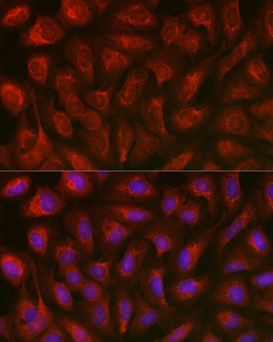
Immunofluorescence analysis of U2OS cells using TXNIP Rabbit pAb (CAB9342) at dilution of 1:100. Secondary antibody: Cy3-conjugated Goat anti-Rabbit IgG (H+L) (CABS007) at 1:500 dilution. Blue: DAPI for nuclear staining.
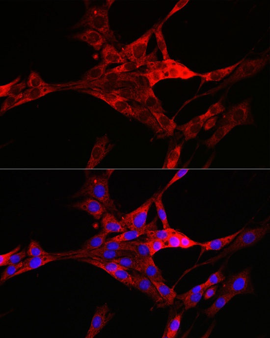
Immunofluorescence analysis of PC-12 cells using TXNIP Rabbit pAb (CAB9342) at dilution of 1:100. Secondary antibody: Cy3-conjugated Goat anti-Rabbit IgG (H+L) (CABS007) at 1:500 dilution. Blue: DAPI for nuclear staining.