The TYR Antibody (CAB13608) is a high-quality antibody developed for reliable detection and analysis of target proteins. This antibody, raised in rabbits, exhibits high reactivity with human samples and has been validated for use in Western blot applications. It specifically binds to the TYR protein, allowing for accurate detection and analysis in a variety of cell types.Tyrosinase is a critical enzyme in the melanin production process, making the TYR gene a key target for research in pigmentation disorders, skin cancer, and other melanin-related conditions.
This antibody is validated for use in WB, IHC-P, ELISA applications and has demonstrated reactivity against Human, Rat samples.
Product Name:
TYR Antibody
SKU:
CAB13608
Size:
20μL, 100μL
Reactivity:
Human, Rat
Conjugate:
Unconjugated
Immunogen:
Recombinant protein (or fragment).This information is considered to be commercially sensitive.
Recommended starting concentration is 1 μg/mL. Please optimize the concentration based on your specific assay requirements.
Synonyms:
ATN, CMM8, OCA1, OCA1A, OCAIA, SHEP3, Tyrosinase
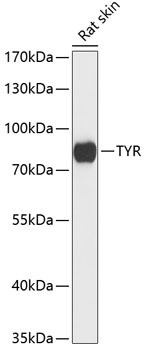
Positive Sample:
Rat skin
Cellular Localization:
Melanosome Membrane, Single-Pass Type I Membrane Protein.
Calculated MW:
60kDa
Observed MW:
80kDa
The enzyme encoded by this gene catalyzes the first 2 steps, and at least 1 subsequent step, in the conversion of tyrosine to melanin. The enzyme has both tyrosine hydroxylase and dopa oxidase catalytic activities, and requires copper for function. Mutations in this gene result in oculocutaneous albinism, and nonpathologic polymorphisms result in skin pigmentation variation. The human genome contains a pseudogene similar to the 3' half of this gene.
Purification Method
Affinity purification
Gene ID
7299
RRID
AB_2760470
Buffer Information
Store at -20℃. Avoid freeze / thaw cycles. Buffer: Buffer: PBS containing 50% glycerol, preserved with proclin300 or sodium azide, pH 7.3.
Western blot analysis of lysates from rat skin, using Tyrosinase Rabbit pAb (CAB13608) at 1:3000 dilution. Secondary antibody: HRP-conjugated Goat anti-Rabbit IgG (H+L) (CABS014) at 1:10000 dilution. Lysates/proteins: 25μg per lane. Blocking buffer: 3% nonfat dry milk in TBST. Detection: ECL Basic Kit (AbGn00020). Exposure time: 10s.
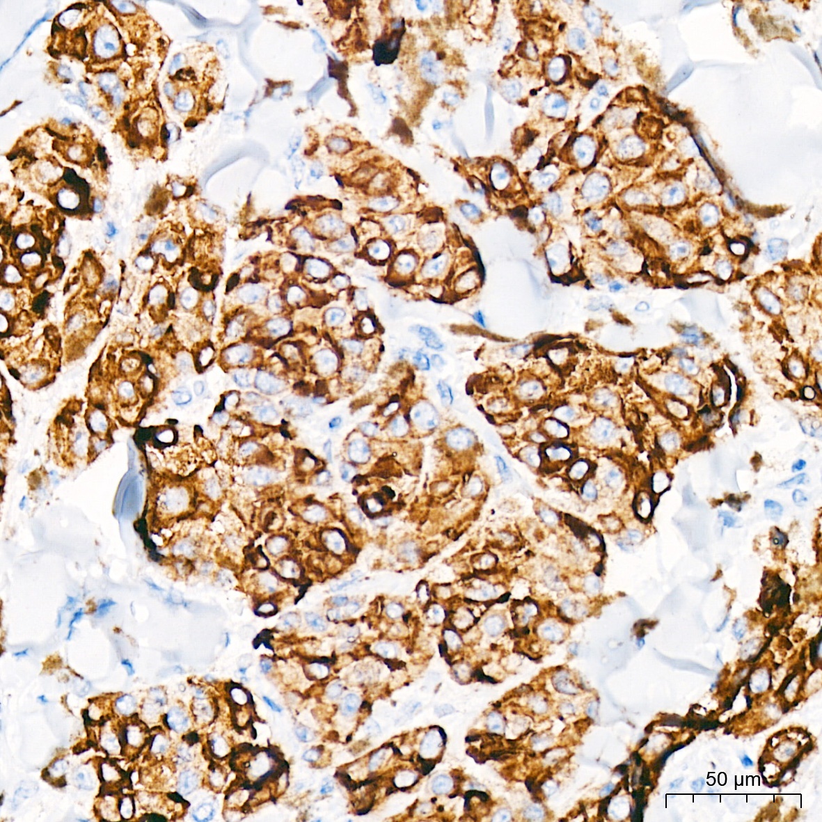
Immunohistochemistry analysis of paraffin-embedded Human malignant melanoma tissue using Tyrosinase Rabbit pAb (CAB13608) at a dilution of 1:2000 (40x lens). High pressure antigen retrieval performed with 0.01M Citrate buffer (pH 6.0) prior to IHC staining.