The UBASH3B Antibody (CAB7141) is a high-quality antibody developed for reliable detection and analysis of target proteins. This antibody, raised in rabbits, exhibits high reactivity with human samples and is validated for use in Western blot applications. By binding to the UBASH3B protein, this antibody enables accurate detection and analysis in a variety of cell types, making it perfect for studies in immunology and cancer research.UBASH3B, also known as T-cell ubiquitin ligand, plays a crucial role in immune responses by regulating signaling pathways and modulating immune cell functions.
This antibody is validated for use in WB, IHC-P, IF/ICC, ELISA applications and has demonstrated reactivity against Human, Mouse, Rat samples.
Product Name:
UBASH3B Antibody
SKU:
CAB7141
Size:
20μL, 100μL
Reactivity:
Human, Mouse, Rat
Conjugate:
Unconjugated
Immunogen:
Recombinant protein (or fragment).This information is considered to be commercially sensitive.
Recommended starting concentration is 1 μg/mL. Please optimize the concentration based on your specific assay requirements.
Synonyms:
p70, STS1, STS-1, TULA2, TULA-2, UBASH3B
Positive Sample:
SW620, SKOV3, Mouse brain, Mouse spleen, Mouse lung, Rat brain
Cellular Localization:
Cytoplasm, Nucleus.
Calculated MW:
73kDa
Observed MW:
72kDa
This gene encodes a protein that contains a ubiquitin associated domain at the N-terminus, an SH3 domain, and a C-terminal domain with similarities to the catalytic motif of phosphoglycerate mutase. The encoded protein was found to inhibit endocytosis of epidermal growth factor receptor (EGFR) and platelet-derived growth factor receptor.
Purification Method
Affinity purification
Gene ID
84959
RRID
AB_2767696
Buffer Information
Store at -20℃. Avoid freeze / thaw cycles. Buffer: PBS containing 50% glycerol, preserved with proclin300 or sodium azide, pH 7.3.
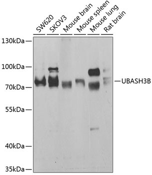
Western blot analysis of various lysates using UBASH3B Rabbit pAb (CAB7141) at 1:1000 dilution. Secondary antibody: HRP-conjugated Goat anti-Rabbit IgG (H+L) (CABS014) at 1:10000 dilution. Lysates/proteins: 25μg per lane. Blocking buffer: 3% nonfat dry milk in TBST. Detection: ECL Basic Kit (AbGn00020). Exposure time: 30s.
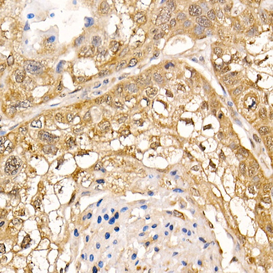
Immunohistochemistry analysis of paraffin-embedded Human lung cancer using UBASH3B Rabbit pAb (CAB7141) at dilution of 1:200 (40x lens). High pressure antigen retrieval performed with 0.01M Citrate buffer (pH 6.0) prior to IHC staining.
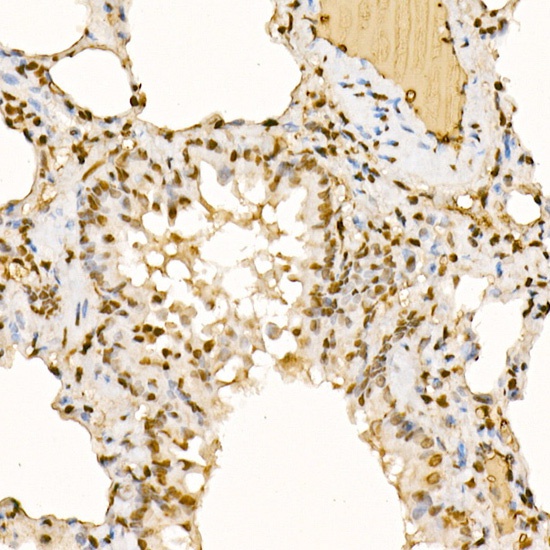
Immunohistochemistry analysis of paraffin-embedded Rat lung using UBASH3B Rabbit pAb (CAB7141) at dilution of 1:200 (40x lens). High pressure antigen retrieval performed with 0.01M Citrate buffer (pH 6.0) prior to IHC staining.
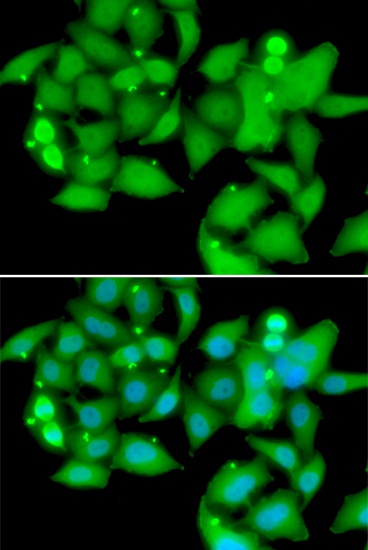
Immunofluorescence analysis of A549 cells using UBASH3B Rabbit pAb (CAB7141). Secondary antibody: Cy3-conjugated Goat anti-Rabbit IgG (H+L) (CABS007) at 1:500 dilution. Blue: DAPI for nuclear staining.