The UBC Antibody (CAB3207) is a high-quality antibody developed for reliable detection and analysis of target proteins. This antibody, produced in rabbits, demonstrates high reactivity with human samples and has been validated for use in Western blot applications. By binding specifically to polyubiquitin C, it enables the detection and analysis of this important protein modification in various cell types.Polyubiquitin C is a key player in the ubiquitin-proteasome system, which controls the degradation of misfolded or damaged proteins, as well as the regulation of protein levels in response to cellular signals.
This antibody is validated for use in WB, IHC-P, IF/ICC, ELISA applications and has demonstrated reactivity against Human, Mouse, Rat samples.
Product Name:
UBC Antibody
SKU:
CAB3207
Size:
20μL, 100μL
Reactivity:
Human, Mouse, Rat
Conjugate:
Unconjugated
Immunogen:
Synthetic peptide. This information is considered to be commercially sensitive.
Recommended starting concentration is 1 μg/mL. Please optimize the concentration based on your specific assay requirements.
Synonyms:
HMG20, UBC
Positive Sample:
293T, BT-474, HepG2, U-251MG, A-549, Mouse testis, Mouse liver, Rat testis, Rat brain, 293F, Hep G2, Mouse testis, Mouse liver, Rat testis
Cellular Localization:
Cytoplasm, Nucleus.
Calculated MW:
77kDa
Observed MW:
12-55kDa/11-77kDa
This gene represents a ubiquitin gene, ubiquitin C. The encoded protein is a polyubiquitin precursor. Conjugation of ubiquitin monomers or polymers can lead to various effects within a cell, depending on the residues to which ubiquitin is conjugated. Ubiquitination has been associated with protein degradation, DNA repair, cell cycle regulation, kinase modification, endocytosis, and regulation of other cell signaling pathways.
Purification Method
Affinity purification
Gene ID
7316
RRID
AB_2764989
Buffer Information
Store at -20℃. Avoid freeze / thaw cycles. Buffer: PBS containing 50% glycerol, preserved with proclin300 or sodium azide, pH 7.3.
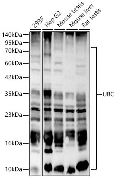
Western blot analysis of various lysates using UBC Rabbit pAb (CAB3207) at 1:1000 dilution. Secondary antibody: HRP-conjugated Goat anti-Rabbit IgG (H+L) (CABS014) at 1:10000 dilution. Lysates / proteins: 25 μg per lane. Blocking buffer: 3 % nonfat dry milk in TBST. Detection: ECL Basic Kit (AbGn00020). Exposure time: 45s.
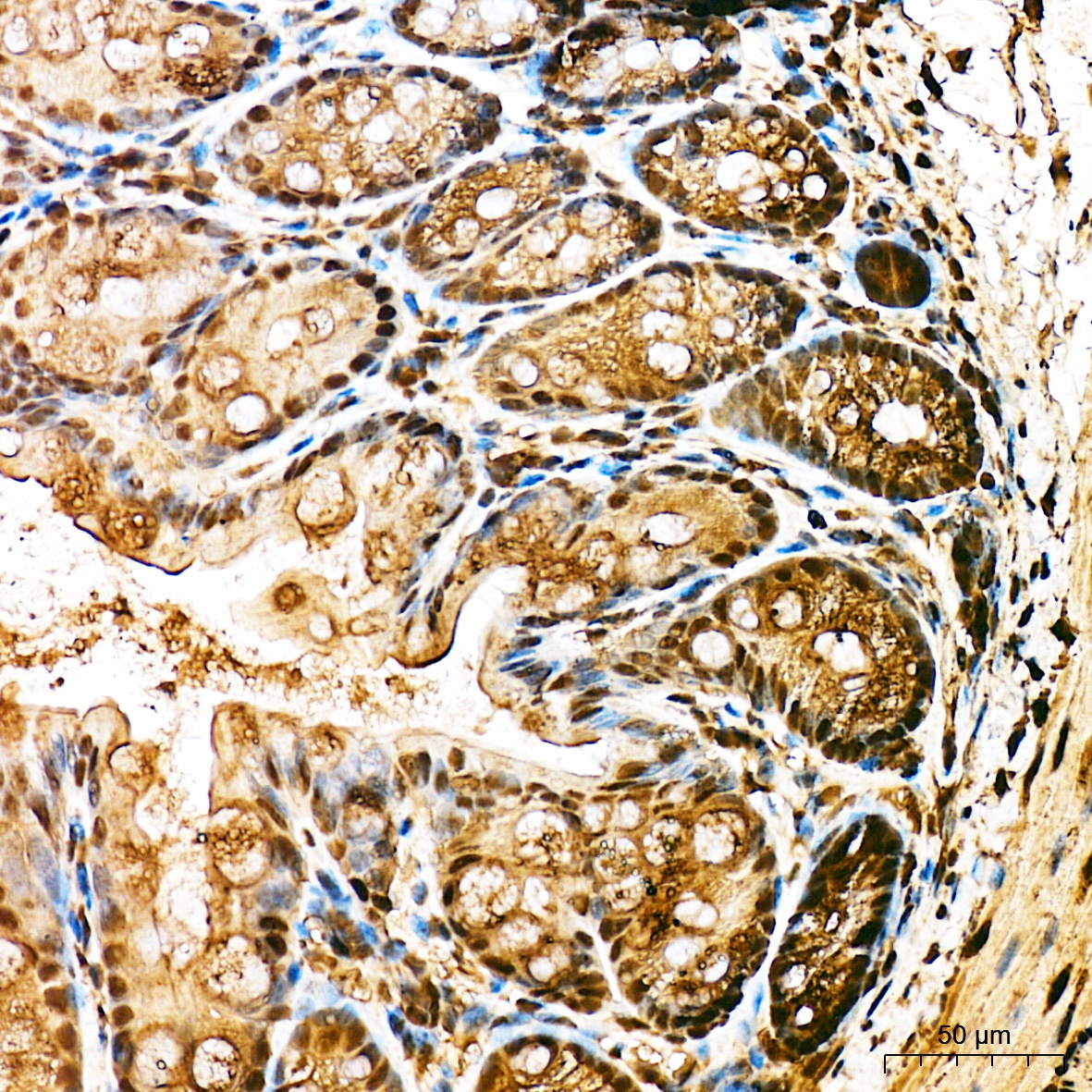
Immunohistochemistry analysis of paraffin-embedded Mouse large intestine tissue using UBC Rabbit pAb (CAB3207) at a dilution of 1:100 (40x lens). High pressure antigen retrieval was performed with 0.01 M citrate buffer (pH 6.0) prior to IHC staining.
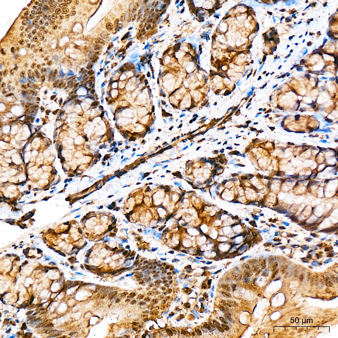
Immunohistochemistry analysis of paraffin-embedded Rat large intestine tissue using UBC Rabbit pAb (CAB3207) at a dilution of 1:100 (40x lens). High pressure antigen retrieval was performed with 0.01 M citrate buffer (pH 6.0) prior to IHC staining.
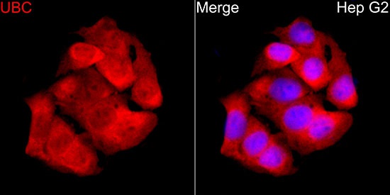
Immunofluorescence analysis of HepG2 cells using UBC Rabbit pAb (CAB3207) at dilution of 1:100 (40x lens). Secondary antibody: Cy3-conjugated Goat anti-Rabbit IgG (H+L) (CABS007) at 1:500 dilution. Blue: DAPI for nuclear staining.